6FPI

Structure of fully reduced Hydrogenase (Hyd-1) variant E28Q


Experimental Data Snapshot

  • Method: X-RAY DIFFRACTION
  • Resolution: 1.50 Å
  • R-Value Free: 0.167 
  • R-Value Work: 0.145 
  • R-Value Observed: 0.146 

wwPDB Validation   3D Report Full Report


Ligand Structure Quality Assessment 


This is version 2.1 of the entry. See complete history


Literature

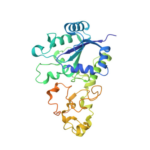
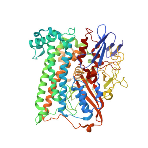
Mechanistic Exploitation of a Self-Repairing, Blocked Proton Transfer Pathway in an O2-Tolerant [NiFe]-Hydrogenase.

Evans, R.M.Ash, P.A.Beaton, S.E.Brooke, E.J.Vincent, K.A.Carr, S.B.Armstrong, F.A.

(2018) J Am Chem Soc 140: 10208-10220

  • DOI: https://doi.org/10.1021/jacs.8b04798
  • Primary Citation of Related Structures:  
    5LRY, 6FPI, 6FPO, 6FPW, 6G7R, 6GAL, 6GAM, 6GAN

  • PubMed Abstract: 

    Catalytic long-range proton transfer in [NiFe]-hydrogenases has long been associated with a highly conserved glutamate (E) situated within 4 Å of the active site. Substituting for glutamine (Q) in the O 2 -tolerant [NiFe]-hydrogenase-1 from Escherichia coli produces a variant (E28Q) with unique properties that have been investigated using protein film electrochemistry, protein film infrared electrochemistry, and X-ray crystallography. At pH 7 and moderate potential, E28Q displays approximately 1% of the activity of the native enzyme, high enough to allow detailed infrared measurements under steady-state conditions. Atomic-level crystal structures reveal partial displacement of the amide side chain by a hydroxide ion, the occupancy of which increases with pH or under oxidizing conditions supporting formation of the superoxidized state of the unusual proximal [4Fe-3S] cluster located nearby. Under these special conditions, the essential exit pathway for at least one of the H + ions produced by H 2 oxidation, and assumed to be blocked in the E28Q variant, is partially repaired. During steady-state H 2 oxidation at neutral pH (i.e., when the barrier to H + exit via Q28 is almost totally closed), the catalytic cycle is dominated by the reduced states "Ni a -R" and "Ni a -C", even under highly oxidizing conditions. Hence, E28 is not involved in the initial activation/deprotonation of H 2 , but facilitates H + exit later in the catalytic cycle to regenerate the initial oxidized active state, assumed to be Ni a -SI. Accordingly, the oxidized inactive resting state, "Ni-B", is not produced by E28Q in the presence of H 2 at high potential because Ni a -SI (the precursor for Ni-B) cannot accumulate. The results have important implications for understanding the catalytic mechanism of [NiFe]-hydrogenases and the control of long-range proton-coupled electron transfer in hydrogenases and other enzymes.


  • Organizational Affiliation

    Department of Chemistry , University of Oxford , Oxford OX1 3QR , United Kingdom.


Macromolecules
Find similar proteins by:  (by identity cutoff)  |  3D Structure
Entity ID: 1
MoleculeChains Sequence LengthOrganismDetailsImage
Hydrogenase-1 small chainA [auth S],
C [auth T]
327Escherichia coli K-12Mutation(s): 0 
EC: 1.12.99.6
UniProt
Find proteins for P69739 (Escherichia coli (strain K12))
Explore P69739 
Go to UniProtKB:  P69739
Entity Groups  
Sequence Clusters30% Identity50% Identity70% Identity90% Identity95% Identity100% Identity
UniProt GroupP69739
Sequence Annotations
Expand
  • Reference Sequence
Find similar proteins by:  (by identity cutoff)  |  3D Structure
Entity ID: 2
MoleculeChains Sequence LengthOrganismDetailsImage
Hydrogenase-1 large chainB [auth L],
D [auth M]
582Escherichia coli K-12Mutation(s): 0 
EC: 1.12.99.6
UniProt
Find proteins for P0ACD8 (Escherichia coli (strain K12))
Explore P0ACD8 
Go to UniProtKB:  P0ACD8
Entity Groups  
Sequence Clusters30% Identity50% Identity70% Identity90% Identity95% Identity100% Identity
UniProt GroupP0ACD8
Sequence Annotations
Expand
  • Reference Sequence
Small Molecules
Ligands 8 Unique
IDChains Name / Formula / InChI Key2D Diagram3D Interactions
LMT
Query on LMT

Download Ideal Coordinates CCD File 
H [auth S],
R [auth T]
DODECYL-BETA-D-MALTOSIDE
C24 H46 O11
NLEBIOOXCVAHBD-QKMCSOCLSA-N
SF4
Query on SF4

Download Ideal Coordinates CCD File 
E [auth S],
O [auth T]
IRON/SULFUR CLUSTER
Fe4 S4
LJBDFODJNLIPKO-UHFFFAOYSA-N
SF3
Query on SF3

Download Ideal Coordinates CCD File 
G [auth S],
Q [auth T]
FE4-S3 CLUSTER
Fe4 S3
QQACTBFBZNWJMV-UHFFFAOYSA-N
F3S
Query on F3S

Download Ideal Coordinates CCD File 
F [auth S],
P [auth T]
FE3-S4 CLUSTER
Fe3 S4
FCXHZBQOKRZXKS-UHFFFAOYSA-N
EJ2
Query on EJ2

Download Ideal Coordinates CCD File 
L,
V [auth M]
NI-FE REDUCED ACTIVE CENTER
C3 H Fe N2 Ni O
JIXQCBCEPWXSQU-UHFFFAOYSA-O
SO4
Query on SO4

Download Ideal Coordinates CCD File 
N [auth L]SULFATE ION
O4 S
QAOWNCQODCNURD-UHFFFAOYSA-L
CL
Query on CL

Download Ideal Coordinates CCD File 
I [auth S]
J [auth S]
K [auth S]
S [auth T]
T
I [auth S],
J [auth S],
K [auth S],
S [auth T],
T,
U [auth T]
CHLORIDE ION
Cl
VEXZGXHMUGYJMC-UHFFFAOYSA-M
MG
Query on MG

Download Ideal Coordinates CCD File 
M [auth L],
W [auth M]
MAGNESIUM ION
Mg
JLVVSXFLKOJNIY-UHFFFAOYSA-N
Experimental Data & Validation

Experimental Data

  • Method: X-RAY DIFFRACTION
  • Resolution: 1.50 Å
  • R-Value Free: 0.167 
  • R-Value Work: 0.145 
  • R-Value Observed: 0.146 
  • Space Group: P 21 21 21
Unit Cell:
Length ( Å )Angle ( ˚ )
a = 93.82α = 90
b = 97.73β = 90
c = 183.2γ = 90
Software Package:
Software NamePurpose
REFMACrefinement
DIALSdata reduction
Aimlessdata scaling
PHASERphasing

Structure Validation

View Full Validation Report


Ligand Structure Quality Assessment 


Entry History & Funding Information

Deposition Data


Funding OrganizationLocationGrant Number
Biotechnology and Biological Sciences Research CouncilUnited KingdomBB/N006321/1

Revision History  (Full details and data files)

  • Version 1.0: 2018-12-19
    Type: Initial release
  • Version 2.0: 2019-01-30
    Changes: Atomic model, Data collection, Derived calculations
  • Version 2.1: 2024-01-17
    Changes: Data collection, Database references, Derived calculations, Refinement description