6DWN

Structure of Human Cytochrome P450 1A1 with Erlotinib

  • Classification: OXIDOREDUCTASE
  • Organism(s): Homo sapiens
  • Expression System: Escherichia coli
  • Mutation(s): No 

  • Deposited: 2018-06-26 Released: 2018-10-03 
  • Deposition Author(s): Bart, A.G., Scott, E.E.
  • Funding Organization(s): National Institutes of Health/National Institute of General Medical Sciences (NIH/NIGMS)

Experimental Data Snapshot

  • Method: X-RAY DIFFRACTION
  • Resolution: 3.00 Å
  • R-Value Free: 0.261 
  • R-Value Work: 0.241 
  • R-Value Observed: 0.242 

wwPDB Validation   3D Report Full Report


Ligand Structure Quality Assessment 


This is version 1.4 of the entry. See complete history


Literature

Structures of human cytochrome P450 1A1 with bergamottin and erlotinib reveal active-site modifications for binding of diverse ligands.

Bart, A.G.Scott, E.E.

(2018) J Biol Chem 293: 19201-19210

  • DOI: https://doi.org/10.1074/jbc.RA118.005588
  • Primary Citation of Related Structures:  
    6DWM, 6DWN

  • PubMed Abstract: 

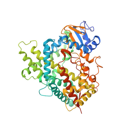
    Human cytochrome P450 1A1 (CYP1A1) is an extrahepatic enzyme involved in the monooxygenation of structurally diverse compounds ranging from natural products to drugs and protoxins. Because CYP1A1 has a role in human carcinogenesis, inhibiting its activity may potentially aid in cancer chemoprevention, whereas utilizing CYP1A1's oxidative activity could help selectively activate anticancer prodrugs. Such potential therapeutic purposes require detailed knowledge of CYP1A1's interactions with potential ligands. Known CYP1A1 ligands also vary substantially in size, and it has not been apparent from a single existing CYP1A1 structure how larger, structurally diverse ligands are accommodated within the enclosed active site. Here, two new X-ray structures with the natural product furanocoumarin bergamottin (at 2.85 Å resolution) and the lung cancer drug erlotinib (3.0 Å) revealed binding orientations consistent with the formation of innocuous metabolites and of toxic metabolites, respectively. They also disclosed local changes in the roof of the active site that enlarge the active site and ultimately form a channel to the protein exterior. Although further structural modifications would be required to accommodate the largest CYP1A1 ligands, knowing which components of the active site are malleable provides powerful information for those attempting to use computational approaches to predict compound binding and substrate metabolism by this clinically relevant monooxygenase.


  • Organizational Affiliation

    From the Program in Biophysics and.


Macromolecules
Find similar proteins by:  (by identity cutoff)  |  3D Structure
Entity ID: 1
MoleculeChains Sequence LengthOrganismDetailsImage
Cytochrome P450 1A1
A, B, C, D
491Homo sapiensMutation(s): 0 
Gene Names: CYP1A1
EC: 1.14.14.1
UniProt & NIH Common Fund Data Resources
Find proteins for P04798 (Homo sapiens)
Explore P04798 
Go to UniProtKB:  P04798
PHAROS:  P04798
GTEx:  ENSG00000140465 
Entity Groups  
Sequence Clusters30% Identity50% Identity70% Identity90% Identity95% Identity100% Identity
UniProt GroupP04798
Sequence Annotations
Expand
  • Reference Sequence
Small Molecules
Ligands 3 Unique
IDChains Name / Formula / InChI Key2D Diagram3D Interactions
HEM
Query on HEM

Download Ideal Coordinates CCD File 
E [auth A],
H [auth B],
K [auth C],
M [auth D]
PROTOPORPHYRIN IX CONTAINING FE
C34 H32 Fe N4 O4
KABFMIBPWCXCRK-RGGAHWMASA-L
CPS
Query on CPS

Download Ideal Coordinates CCD File 
G [auth A],
J [auth B]
3-[(3-CHOLAMIDOPROPYL)DIMETHYLAMMONIO]-1-PROPANESULFONATE
C32 H58 N2 O7 S
UMCMPZBLKLEWAF-BCTGSCMUSA-N
AQ4
Query on AQ4

Download Ideal Coordinates CCD File 
F [auth A],
I [auth B],
L [auth C],
N [auth D]
[6,7-BIS(2-METHOXY-ETHOXY)QUINAZOLINE-4-YL]-(3-ETHYNYLPHENYL)AMINE
C22 H23 N3 O4
AAKJLRGGTJKAMG-UHFFFAOYSA-N
Experimental Data & Validation

Experimental Data

  • Method: X-RAY DIFFRACTION
  • Resolution: 3.00 Å
  • R-Value Free: 0.261 
  • R-Value Work: 0.241 
  • R-Value Observed: 0.242 
  • Space Group: P 21 21 21
Unit Cell:
Length ( Å )Angle ( ˚ )
a = 64.855α = 90
b = 195.078β = 90
c = 238.013γ = 90
Software Package:
Software NamePurpose
PHENIXrefinement
XDSdata reduction
Aimlessdata scaling
PHASERphasing

Structure Validation

View Full Validation Report


Ligand Structure Quality Assessment 


Entry History & Funding Information

Deposition Data


Funding OrganizationLocationGrant Number
National Institutes of Health/National Institute of General Medical Sciences (NIH/NIGMS)United StatesGM076343

Revision History  (Full details and data files)

  • Version 1.0: 2018-10-03
    Type: Initial release
  • Version 1.1: 2018-10-10
    Changes: Data collection, Database references, Structure summary
  • Version 1.2: 2018-12-26
    Changes: Data collection, Database references, Structure summary
  • Version 1.3: 2020-01-01
    Changes: Author supporting evidence
  • Version 1.4: 2023-10-11
    Changes: Data collection, Database references, Refinement description