6AE3

Crystal structure of GSK3beta complexed with Morin


Experimental Data Snapshot

  • Method: X-RAY DIFFRACTION
  • Resolution: 2.14 Å
  • R-Value Free: 0.226 
  • R-Value Work: 0.191 
  • R-Value Observed: 0.192 

wwPDB Validation   3D Report Full Report


Ligand Structure Quality Assessment 


This is version 1.2 of the entry. See complete history


Literature

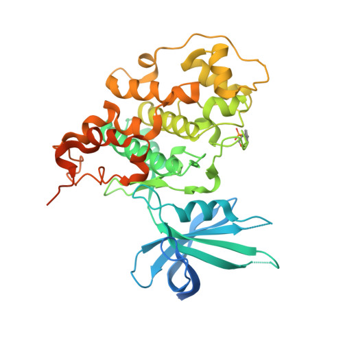
Crystal structure of GSK3 beta in complex with the flavonoid, morin

Kim, K.Cha, J.S.Kim, J.S.Ahn, J.Ha, N.C.Cho, H.S.

(2018) Biochem Biophys Res Commun 504: 519-524

  • DOI: https://doi.org/10.1016/j.bbrc.2018.08.182
  • Primary Citation of Related Structures:  
    6AE3

  • PubMed Abstract: 

    GSK3β is a key kinase that plays a role in cellular signaling pathways. In Alzheimer's disease (AD), GSK3β has been implicated in hyperphosphorylation of tau proteins in the neuron, which is a hallmark of AD. Morin, a flavonoid that is abundant in nature, was found as an inhibitor of GSK3β that can reduce tau pathology in vivo and in vitro. In this study, we determined the crystal structure of GSK3β in complex with morin. The structure revealed that morin inhibits GSK3β by binding to the ATP binding pocket. Our findings augment the potential of morin as a functional food to help prevent AD, as well as to provide structural information to develop new therapeutics based on the morin skeleton.


  • Organizational Affiliation

    Department of Systems Biology, College of Life Science and Biotechnology, Yonsei University, Seoul 120-749, Republic of Korea; Department of Pharmacology, University of North Carolina at Chapel Hill School of Medicine, Chapel Hill, NC 27516, USA.


Macromolecules
Find similar proteins by:  (by identity cutoff)  |  3D Structure
Entity ID: 1
MoleculeChains Sequence LengthOrganismDetailsImage
Glycogen synthase kinase-3 beta
A, B, C, D
420Mus musculusMutation(s): 0 
Gene Names: Gsk3b
EC: 2.7.11.26 (PDB Primary Data), 2.7.11.1 (PDB Primary Data)
UniProt
Find proteins for Q9WV60 (Mus musculus)
Explore Q9WV60 
Go to UniProtKB:  Q9WV60
Entity Groups  
Sequence Clusters30% Identity50% Identity70% Identity90% Identity95% Identity100% Identity
UniProt GroupQ9WV60
Sequence Annotations
Expand
  • Reference Sequence
Small Molecules
Experimental Data & Validation

Experimental Data

  • Method: X-RAY DIFFRACTION
  • Resolution: 2.14 Å
  • R-Value Free: 0.226 
  • R-Value Work: 0.191 
  • R-Value Observed: 0.192 
  • Space Group: P 1 21 1
Unit Cell:
Length ( Å )Angle ( ˚ )
a = 67.588α = 90
b = 134.36β = 103.8
c = 100.393γ = 90
Software Package:
Software NamePurpose
REFMACrefinement
HKL-2000data reduction
HKL-2000data scaling
MOLREPphasing

Structure Validation

View Full Validation Report


Ligand Structure Quality Assessment 


Entry History 

Deposition Data

Revision History  (Full details and data files)

  • Version 1.0: 2018-09-19
    Type: Initial release
  • Version 1.1: 2018-09-26
    Changes: Data collection, Database references
  • Version 1.2: 2018-10-03
    Changes: Data collection, Database references