6A12

X-ray structure of lipase from Geobacillus thermoleovorans


Experimental Data Snapshot

  • Method: X-RAY DIFFRACTION
  • Resolution: 2.15 Å
  • R-Value Free: 0.221 
  • R-Value Work: 0.173 
  • R-Value Observed: 0.175 

wwPDB Validation   3D Report Full Report


This is version 1.2 of the entry. See complete history


Literature

X-ray structure and characterization of a thermostable lipase from Geobacillus thermoleovorans.

Moharana, T.R.Pal, B.Rao, N.M.

(2019) Biochem Biophys Res Commun 508: 145-151

  • DOI: https://doi.org/10.1016/j.bbrc.2018.11.105
  • Primary Citation of Related Structures:  
    6A12

  • PubMed Abstract: 

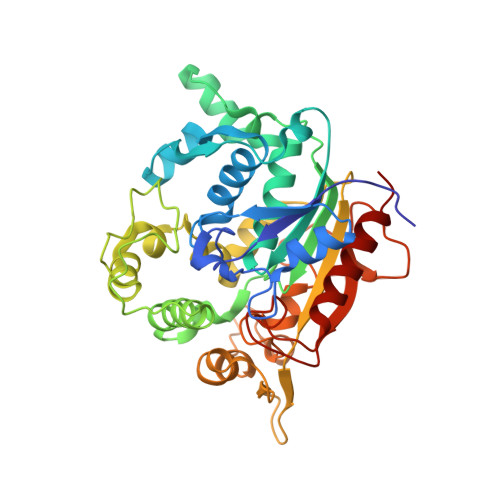
    Thermo-alkalophilic bacterium, Geobacillus thermoleovorans secrets many enzymes including a 43 kDa extracellular lipase. Significant thermostability, organic solvent stability and wide substrate preferences for hydrolysis drew our attention to solve its structure by crystallography. The structure was solved by molecular replacement method and refined up to 2.14 Å resolution. Structure of the lipase showed an alpha-beta fold with 19 α-helices and 10 β-sheets. The active site remains covered by a lid. One calcium and one zinc atom was found in the crystal. The structure showed a major difference (rmsd 5.6 Å) from its closest homolog in the amino acid region 191 to 203. Thermal unfolding of the lipase showed that the lipase is highly stable with T m of 76 °C. 13 C NMR spectra of products upon triglyceride hydrolysate revealed that the lipase hydrolyses at both sn-1 and sn-2 positions with equal efficiency.


  • Organizational Affiliation

    CSIR- Centre for Cellular and Molecular Biology, Uppal Road, Hyderabad, 500007, India.


Macromolecules
Find similar proteins by:  (by identity cutoff)  |  3D Structure
Entity ID: 1
MoleculeChains Sequence LengthOrganismDetailsImage
Lipase389Geobacillus thermoleovoransMutation(s): 0 
Gene Names: lipA
EC: 3.1.1.3
UniProt
Find proteins for Q8L1V2 (Geobacillus thermoleovorans)
Explore Q8L1V2 
Go to UniProtKB:  Q8L1V2
Entity Groups  
Sequence Clusters30% Identity50% Identity70% Identity90% Identity95% Identity100% Identity
UniProt GroupQ8L1V2
Sequence Annotations
Expand
  • Reference Sequence
Experimental Data & Validation

Experimental Data

  • Method: X-RAY DIFFRACTION
  • Resolution: 2.15 Å
  • R-Value Free: 0.221 
  • R-Value Work: 0.173 
  • R-Value Observed: 0.175 
  • Space Group: P 21 21 21
Unit Cell:
Length ( Å )Angle ( ˚ )
a = 49.14α = 90
b = 72.291β = 90
c = 112.405γ = 90
Software Package:
Software NamePurpose
PHENIXrefinement
HKL-2000data reduction
HKL-2000data scaling
MOLREPphasing

Structure Validation

View Full Validation Report


Entry History 

Deposition Data

Revision History  (Full details and data files)

  • Version 1.0: 2018-06-20
    Type: Initial release
  • Version 1.1: 2019-01-16
    Changes: Data collection, Database references
  • Version 1.2: 2023-11-22
    Changes: Data collection, Database references, Refinement description