5ZIQ

Crystal structure of hexacoordinated heme protein from anhydrobiotic tardigrade at pH 4


Experimental Data Snapshot

  • Method: X-RAY DIFFRACTION
  • Resolution: 1.50 Å
  • R-Value Free: 0.194 
  • R-Value Work: 0.147 
  • R-Value Observed: 0.149 

wwPDB Validation   3D Report Full Report


Ligand Structure Quality Assessment 


This is version 1.2 of the entry. See complete history


Literature

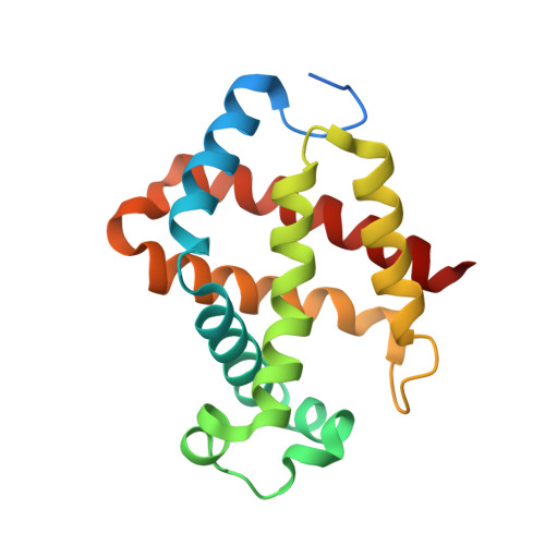
Crystal structure of Kumaglobin: a hexacoordinated heme protein from an anhydrobiotic tardigrade, Ramazzottius varieornatus.

Kim, J.Fukuda, Y.Inoue, T.

(2019) FEBS J 286: 1287-1304

  • DOI: https://doi.org/10.1111/febs.14713
  • Primary Citation of Related Structures:  
    5ZIQ, 5ZM9

  • PubMed Abstract: 

    Tardigrades, also known as water bears, can survive extreme conditions. For example, tardigrades have high tolerance to extreme desiccation because they can enter an anhydrobiotic state, in which they show no or nearly undetectable metabolic processes. Proteins from anhydrobiotic tardigrades with low homology to known proteins from other organisms are new potential targets for structural genomics. Here, we present spectroscopic and structural characterization of an unprecedented globin protein (Kumaglobin: Kgb) found in an anhydrobiotic tardigrade. Spectroscopy reveals that Kgb contains hexacoordinated low-spin heme, which is not capable of binding to hydrogen sulfide (H 2 S) unlike other globin proteins, such as neuroglobin. Interestingly however, when distal histidine is replaced with alanine, H 2 S is capable of binding to heme, implying that the distal histidine of Kgb binds tightly to heme. The overall structure of Kgb at 1.5 Å resolution shows high resemblance to well-characterized eukaryotic globin proteins, such as myoglobin and cytoglobin. However, the heme coordination geometry in Kgb is unique because the distal histidinyl ligand is located at the 11th position of helix E while it is found at 7th position on helix E in many known globin proteins. The unusual conformation of distal histidine in Kgb is stabilized by a hydrogen bond with the carbonyl O atom of A103. Furthermore, bulky residues exist around the heme cofactor, resulting in a ruffling conformation of the porphyrin ring. Based on our study, Kgb is thought to be involved in electron transfer or enzymatic reactions rather than transporting or storing ligands. DATABASE: Structural data are available in the Protein Data Bank under the accession numbers 5ZIQ (Kgb4-SR) and 5ZM9 (Kgb7-house).


  • Organizational Affiliation

    Department of Applied Chemistry, Graduate School of Engineering, Osaka University, Suita, Japan.


Macromolecules
Find similar proteins by:  (by identity cutoff)  |  3D Structure
Entity ID: 1
MoleculeChains Sequence LengthOrganismDetailsImage
Globin protein
A, B, C, D
171Ramazzottius varieornatusMutation(s): 0 
Gene Names: RvY_09119-1RvY_09119.1RvY_09119
UniProt
Find proteins for A0A1D1V896 (Ramazzottius varieornatus)
Explore A0A1D1V896 
Go to UniProtKB:  A0A1D1V896
Entity Groups  
Sequence Clusters30% Identity50% Identity70% Identity90% Identity95% Identity100% Identity
UniProt GroupA0A1D1V896
Sequence Annotations
Expand
  • Reference Sequence
Small Molecules
Ligands 3 Unique
IDChains Name / Formula / InChI Key2D Diagram3D Interactions
HEM
Query on HEM

Download Ideal Coordinates CCD File 
E [auth A],
J [auth B],
O [auth C],
R [auth D]
PROTOPORPHYRIN IX CONTAINING FE
C34 H32 Fe N4 O4
KABFMIBPWCXCRK-RGGAHWMASA-L
SO4
Query on SO4

Download Ideal Coordinates CCD File 
F [auth A]
G [auth A]
H [auth A]
K [auth B]
L [auth B]
F [auth A],
G [auth A],
H [auth A],
K [auth B],
L [auth B],
M [auth B],
P [auth C]
SULFATE ION
O4 S
QAOWNCQODCNURD-UHFFFAOYSA-L
EDO
Query on EDO

Download Ideal Coordinates CCD File 
I [auth A],
N [auth B],
Q [auth C]
1,2-ETHANEDIOL
C2 H6 O2
LYCAIKOWRPUZTN-UHFFFAOYSA-N
Experimental Data & Validation

Experimental Data

  • Method: X-RAY DIFFRACTION
  • Resolution: 1.50 Å
  • R-Value Free: 0.194 
  • R-Value Work: 0.147 
  • R-Value Observed: 0.149 
  • Space Group: P 1
  • Diffraction Data: https://doi.org/10.18430/m35ziq
Unit Cell:
Length ( Å )Angle ( ˚ )
a = 43.359α = 115.36
b = 59.185β = 97.98
c = 67.521γ = 97.43
Software Package:
Software NamePurpose
REFMACrefinement
HKL-2000data reduction
HKL-2000data scaling
MOLREPphasing

Structure Validation

View Full Validation Report


Ligand Structure Quality Assessment 


Entry History & Funding Information

Deposition Data


Funding OrganizationLocationGrant Number
JSPSJapan--

Revision History  (Full details and data files)

  • Version 1.0: 2019-01-02
    Type: Initial release
  • Version 1.1: 2019-04-24
    Changes: Data collection, Database references
  • Version 1.2: 2024-03-27
    Changes: Data collection, Database references