5VBN

Crystal Structure of human DNA polymerase epsilon B-subunit in complex with C-terminal domain of catalytic subunit


Experimental Data Snapshot

  • Method: X-RAY DIFFRACTION
  • Resolution: 2.35 Å
  • R-Value Free: 0.264 
  • R-Value Work: 0.225 
  • R-Value Observed: 0.225 

wwPDB Validation   3D Report Full Report


This is version 1.6 of the entry. See complete history


Literature

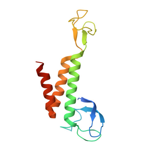
Crystal structure of the human Pol B-subunit in complex with the C-terminal domain of the catalytic subunit.

Baranovskiy, A.G.Gu, J.Babayeva, N.D.Kurinov, I.Pavlov, Y.I.Tahirov, T.H.

(2017) J Biol Chem 292: 15717-15730

  • DOI: https://doi.org/10.1074/jbc.M117.792705
  • Primary Citation of Related Structures:  
    5VBN

  • PubMed Abstract: 

    The eukaryotic B-family DNA polymerases include four members: Polα, Polδ, Polϵ, and Polζ, which share common architectural features, such as the exonuclease/polymerase and C-terminal domains (CTDs) of catalytic subunits bound to indispensable B-subunits, which serve as scaffolds that mediate interactions with other components of the replication machinery. Crystal structures for the B-subunits of Polα and Polδ/Polζ have been reported: the former within the primosome and separately with CTD and the latter with the N-terminal domain of the C-subunit. Here we present the crystal structure of the human Polϵ B-subunit (p59) in complex with CTD of the catalytic subunit (p261 C ). The structure revealed a well defined electron density for p261 C and the phosphodiesterase and oligonucleotide/oligosaccharide-binding domains of p59. However, electron density was missing for the p59 N-terminal domain and for the linker connecting it to the phosphodiesterase domain. Similar to Polα, p261 C of Polϵ contains a three-helix bundle in the middle and zinc-binding modules on each side. Intersubunit interactions involving 11 hydrogen bonds and numerous hydrophobic contacts account for stable complex formation with a buried surface area of 3094 Å 2 Comparative structural analysis of p59-p261 C with the corresponding Polα complex revealed significant differences between the B-subunits and CTDs, as well as their interaction interfaces. The B-subunit of Polδ/Polζ also substantially differs from B-subunits of either Polα or Polϵ. This work provides a structural basis to explain biochemical and genetic data on the importance of B-subunit integrity in replisome function in vivo .


  • Organizational Affiliation

    From the Eppley Institute for Research in Cancer and Allied Diseases, Fred & Pamela Buffett Cancer Center and.


Macromolecules
Find similar proteins by:  (by identity cutoff)  |  3D Structure
Entity ID: 1
MoleculeChains Sequence LengthOrganismDetailsImage
DNA polymerase epsilon subunit 2A,
C [auth E]
527Homo sapiensMutation(s): 0 
Gene Names: POLE2DPE2
EC: 2.7.7.7
UniProt & NIH Common Fund Data Resources
Find proteins for P56282 (Homo sapiens)
Explore P56282 
Go to UniProtKB:  P56282
PHAROS:  P56282
GTEx:  ENSG00000100479 
Entity Groups  
Sequence Clusters30% Identity50% Identity70% Identity90% Identity95% Identity100% Identity
UniProt GroupP56282
Sequence Annotations
Expand
  • Reference Sequence
Find similar proteins by:  (by identity cutoff)  |  3D Structure
Entity ID: 2
MoleculeChains Sequence LengthOrganismDetailsImage
DNA polymerase epsilon catalytic subunit AB,
D [auth F]
145Homo sapiensMutation(s): 0 
Gene Names: POLEPOLE1
EC: 2.7.7.7
UniProt & NIH Common Fund Data Resources
Find proteins for Q07864 (Homo sapiens)
Explore Q07864 
Go to UniProtKB:  Q07864
PHAROS:  Q07864
GTEx:  ENSG00000177084 
Entity Groups  
Sequence Clusters30% Identity50% Identity70% Identity90% Identity95% Identity100% Identity
UniProt GroupQ07864
Sequence Annotations
Expand
  • Reference Sequence
Experimental Data & Validation

Experimental Data

  • Method: X-RAY DIFFRACTION
  • Resolution: 2.35 Å
  • R-Value Free: 0.264 
  • R-Value Work: 0.225 
  • R-Value Observed: 0.225 
  • Space Group: P 21 21 2
Unit Cell:
Length ( Å )Angle ( ˚ )
a = 92.26α = 90
b = 201.459β = 90
c = 78.336γ = 90
Software Package:
Software NamePurpose
CNSrefinement
HKL-2000data reduction
HKL-2000data scaling
PHENIXphasing

Structure Validation

View Full Validation Report


Entry History & Funding Information

Deposition Data


Funding OrganizationLocationGrant Number
National Institutes of Health/National Institute of General Medical Sciences (NIH/NIGMS)United StatesGM101167
National Institutes of Health/National Institute of General Medical Sciences (NIH/NIGMS)United StatesGM103403
National Institutes of Health/National Cancer Institute (NIH/NCI)United StatesCA036727

Revision History  (Full details and data files)

  • Version 1.0: 2017-08-02
    Type: Initial release
  • Version 1.1: 2017-08-09
    Changes: Database references
  • Version 1.2: 2017-09-13
    Changes: Author supporting evidence
  • Version 1.3: 2017-10-04
    Changes: Database references
  • Version 1.4: 2018-10-10
    Changes: Data collection, Refinement description, Structure summary
  • Version 1.5: 2019-12-04
    Changes: Author supporting evidence, Structure summary
  • Version 1.6: 2024-03-06
    Changes: Data collection, Database references