5CW6

Structure of metal dependent enzyme DrBRCC36


Experimental Data Snapshot

  • Method: X-RAY DIFFRACTION
  • Resolution: 3.19 Å
  • R-Value Free: 0.241 
  • R-Value Work: 0.214 
  • R-Value Observed: 0.215 

wwPDB Validation   3D Report Full Report


This is version 1.2 of the entry. See complete history


Literature

Higher-Order Assembly of BRCC36-KIAA0157 Is Required for DUB Activity and Biological Function.

Zeqiraj, E.Tian, L.Piggott, C.A.Pillon, M.C.Duffy, N.M.Ceccarelli, D.F.Keszei, A.F.Lorenzen, K.Kurinov, I.Orlicky, S.Gish, G.D.Heck, A.J.Guarne, A.Greenberg, R.A.Sicheri, F.

(2015) Mol Cell 59: 970-983

  • DOI: https://doi.org/10.1016/j.molcel.2015.07.028
  • Primary Citation of Related Structures:  
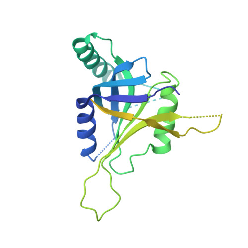
    5CW3, 5CW4, 5CW5, 5CW6

  • PubMed Abstract: 

    BRCC36 is a Zn(2+)-dependent deubiquitinating enzyme (DUB) that hydrolyzes lysine-63-linked ubiquitin chains as part of distinct macromolecular complexes that participate in either interferon signaling or DNA-damage recognition. The MPN(+) domain protein BRCC36 associates with pseudo DUB MPN(-) proteins KIAA0157 or Abraxas, which are essential for BRCC36 enzymatic activity. To understand the basis for BRCC36 regulation, we have solved the structure of an active BRCC36-KIAA0157 heterodimer and an inactive BRCC36 homodimer. Structural and functional characterizations show how BRCC36 is switched to an active conformation by contacts with KIAA0157. Higher-order association of BRCC36 and KIAA0157 into a dimer of heterodimers (super dimers) was required for DUB activity and interaction with targeting proteins SHMT2 and RAP80. These data provide an explanation of how an inactive pseudo DUB allosterically activates a cognate DUB partner and implicates super dimerization as a new regulatory mechanism underlying BRCC36 DUB activity, subcellular localization, and biological function.


  • Organizational Affiliation

    Lunenfeld-Tanenbaum Research Institute, Mount Sinai Hospital, 600 University Avenue, Room 1090, Toronto, ON M5G 1X5, Canada.


Macromolecules
Find similar proteins by:  (by identity cutoff)  |  3D Structure
Entity ID: 1
MoleculeChains Sequence LengthOrganismDetailsImage
DrBRCC36262Danio rerioMutation(s): 0 
UniProt
Find proteins for A0A8M3B525 (Danio rerio)
Explore A0A8M3B525 
Go to UniProtKB:  A0A8M3B525
Entity Groups  
Sequence Clusters30% Identity50% Identity70% Identity90% Identity95% Identity100% Identity
UniProt GroupA0A8M3B525
Sequence Annotations
Expand
  • Reference Sequence
Small Molecules
Ligands 1 Unique
IDChains Name / Formula / InChI Key2D Diagram3D Interactions
ZN
Query on ZN

Download Ideal Coordinates CCD File 
B [auth A]ZINC ION
Zn
PTFCDOFLOPIGGS-UHFFFAOYSA-N
Experimental Data & Validation

Experimental Data

  • Method: X-RAY DIFFRACTION
  • Resolution: 3.19 Å
  • R-Value Free: 0.241 
  • R-Value Work: 0.214 
  • R-Value Observed: 0.215 
  • Space Group: P 62 2 2
Unit Cell:
Length ( Å )Angle ( ˚ )
a = 108.833α = 90
b = 108.833β = 90
c = 137.439γ = 120
Software Package:
Software NamePurpose
PHENIXrefinement
PDB_EXTRACTdata extraction

Structure Validation

View Full Validation Report


Entry History 

Deposition Data

  • Released Date: 2015-09-16 
  • Deposition Author(s): Zeqiraj, E.

Revision History  (Full details and data files)

  • Version 1.0: 2015-09-16
    Type: Initial release
  • Version 1.1: 2015-09-30
    Changes: Database references
  • Version 1.2: 2024-03-06
    Changes: Data collection, Database references, Derived calculations