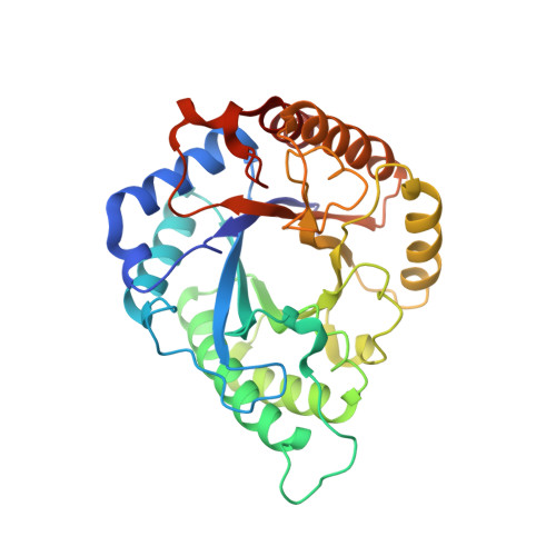
Structural insights into the catalytic mechanism of a novel glycoside hydrolase family 113 beta-1,4-mannanase from Amphibacillus xylanus
You, X., Qin, Z., Yan, Q., Yang, S., Li, Y., Jiang, Z.(2018) J Biol Chem 293: 11746-11757
- PubMed: 29871927
- DOI: https://doi.org/10.1074/jbc.RA118.002363
- Primary Citation of Related Structures:
5YLH, 5YLI, 5YLK, 5YLL, 5Z4T - PubMed Abstract:
β-1,4-Mannanase degrades β-1,4-mannan polymers into manno-oligosaccharides with a low degree of polymerization. To date, only one glycoside hydrolase (GH) family 113 β-1,4-mannanase, from Alicyclobacillus acidocaldarius ( Aa ManA), has been structurally characterized, and no complex structure of enzyme-manno-oligosaccharides from this family has been reported. Here, crystal structures of a GH family 113 β-1,4-mannanase from Amphibacillus xylanus ( Ax Man113A) and its complexes with mannobiose, mannotriose, mannopentaose, and mannahexaose were solved. Ax Man113A had higher affinity for -1 and +1 mannoses, which explains why the enzyme can hydrolyze mannobiose. At least six subsites (-4 to +2) exist in the groove, but mannose units preferentially occupied subsites -4 to -1 because of steric hindrance formed by Lys-238 and Trp-239. Based on the structural information and bioinformatics, rational design was implemented to enhance hydrolysis activity. Enzyme activity of Ax Man113A mutants V139C, N237W, K238A, and W239Y was improved by 93.7, 63.4, 112.9, and 36.4%, respectively, compared with the WT. In addition, previously unreported surface-binding sites were observed. Site-directed mutagenesis studies and kinetic data indicated that key residues near the surface sites play important roles in substrate binding and recognition. These first GH family 113 β-1,4-mannanase-manno-oligosaccharide complex structures may be useful in further studying the catalytic mechanism of GH family 113 members, and provide novel insight into protein engineering of GHs to improve their hydrolysis activity.
Organizational Affiliation:
From the Beijing Advanced Innovation Center for Food Nutrition and Human Health, College of Engineering, China Agricultural University, Beijing 100083.