5XB7

GH42 alpha-L-arabinopyranosidase from Bifidobacterium animalis subsp. lactis Bl-04


Experimental Data Snapshot

  • Method: X-RAY DIFFRACTION
  • Resolution: 2.00 Å
  • R-Value Free: 0.191 
  • R-Value Work: 0.155 
  • R-Value Observed: 0.157 

wwPDB Validation   3D Report Full Report


This is version 1.3 of the entry. See complete history


Literature

Discovery of alpha-l-arabinopyranosidases from human gut microbiome expands the diversity within glycoside hydrolase family 42.

Viborg, A.H.Katayama, T.Arakawa, T.Abou Hachem, M.Lo Leggio, L.Kitaoka, M.Svensson, B.Fushinobu, S.

(2017) J Biol Chem 292: 21092-21101

  • DOI: https://doi.org/10.1074/jbc.M117.792598
  • Primary Citation of Related Structures:  
    5XB7

  • PubMed Abstract: 

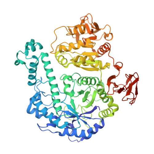
    Enzymes of the glycoside hydrolase family 42 (GH42) are widespread in bacteria of the human gut microbiome and play fundamental roles in the decomposition of both milk and plant oligosaccharides. All GH42 enzymes characterized so far have β-galactosidase activity. Here, we report the existence of a GH42 subfamily that is exclusively specific for α-l-arabinopyranoside and describe the first representative of this subfamily. We found that this enzyme ( Bl Arap42B) from a probiotic Bifidobacterium species cannot hydrolyze β-galactosides. However, Bl Arap42B effectively hydrolyzed paeonolide and ginsenoside Rb2, plant glycosides containing an aromatic aglycone conjugated to α-l-arabinopyranosyl-(1,6)-β-d-glucopyranoside. Paeonolide, a natural glycoside from the roots of the plant genus Paeonia, is not hydrolyzed by classical GH42 β-galactosidases. X-ray crystallography revealed a unique Trp 345 - X 12 -Trp 358 sequence motif at the Bl Arap42B active site, as compared with a Phe- X 12 -His motif in classical GH42 β-galactosidases. This analysis also indicated that the C6 position of galactose is blocked by the aromatic side chains, hence allowing accommodation only of Ara p lacking this carbon. Automated docking of paeonolide revealed that it can fit into the Bl Ara p 42B active site. The Glc p moiety of paeonolide stacks onto the aromatic ring of the Trp 252 at subsite +1 and C4-OH is hydrogen bonded with Asp 249 Moreover, the aglycone stacks against Phe 421 from the neighboring monomer in the Bl Ara p 42B trimer, forming a proposed subsite +2. These results further support the notion that evolution of metabolic specialization can be tracked at the structural level in key enzymes facilitating degradation of specific glycans in an ecological niche.


  • Organizational Affiliation

    From the Department of Biotechnology, The University of Tokyo, Tokyo 113-8657, Japan.


Macromolecules
Find similar proteins by:  (by identity cutoff)  |  3D Structure
Entity ID: 1
MoleculeChains Sequence LengthOrganismDetailsImage
Beta-galactosidase
A, B, C, D, E
A, B, C, D, E, F
712Bifidobacterium animalis subsp. lactisMutation(s): 0 
Gene Names: AL0646_0018
UniProt
Find proteins for A0A2H4A2Z3 (Bifidobacterium animalis subsp. lactis)
Explore A0A2H4A2Z3 
Go to UniProtKB:  A0A2H4A2Z3
Entity Groups  
Sequence Clusters30% Identity50% Identity70% Identity90% Identity95% Identity100% Identity
UniProt GroupA0A2H4A2Z3
Sequence Annotations
Expand
  • Reference Sequence
Small Molecules
Ligands 2 Unique
IDChains Name / Formula / InChI Key2D Diagram3D Interactions
SO4
Query on SO4

Download Ideal Coordinates CCD File 
AB [auth E]
BB [auth E]
FA [auth B]
G [auth A]
GA [auth C]
AB [auth E],
BB [auth E],
FA [auth B],
G [auth A],
GA [auth C],
HA [auth C],
KB [auth E],
LB [auth F],
MB [auth F],
P [auth A],
Q [auth B],
R [auth B],
RA [auth D],
S [auth B],
SA [auth D],
YB [auth F]
SULFATE ION
O4 S
QAOWNCQODCNURD-UHFFFAOYSA-L
GOL
Query on GOL

Download Ideal Coordinates CCD File 
AA [auth B]
BA [auth B]
CA [auth B]
CB [auth E]
DA [auth B]
AA [auth B],
BA [auth B],
CA [auth B],
CB [auth E],
DA [auth B],
DB [auth E],
EA [auth B],
EB [auth E],
FB [auth E],
GB [auth E],
H [auth A],
HB [auth E],
I [auth A],
IA [auth C],
IB [auth E],
J [auth A],
JA [auth C],
JB [auth E],
K [auth A],
KA [auth C],
L [auth A],
LA [auth C],
M [auth A],
MA [auth C],
N [auth A],
NA [auth C],
NB [auth F],
O [auth A],
OA [auth C],
OB [auth F],
PA [auth C],
PB [auth F],
QA [auth C],
QB [auth F],
RB [auth F],
SB [auth F],
T [auth B],
TA [auth D],
TB [auth F],
U [auth B],
UA [auth D],
UB [auth F],
V [auth B],
VA [auth D],
VB [auth F],
W [auth B],
WA [auth D],
WB [auth F],
X [auth B],
XA [auth D],
XB [auth F],
Y [auth B],
YA [auth D],
Z [auth B],
ZA [auth D]
GLYCEROL
C3 H8 O3
PEDCQBHIVMGVHV-UHFFFAOYSA-N
Experimental Data & Validation

Experimental Data

  • Method: X-RAY DIFFRACTION
  • Resolution: 2.00 Å
  • R-Value Free: 0.191 
  • R-Value Work: 0.155 
  • R-Value Observed: 0.157 
  • Space Group: P 41 21 2
Unit Cell:
Length ( Å )Angle ( ˚ )
a = 177.954α = 90
b = 177.954β = 90
c = 375.697γ = 90
Software Package:
Software NamePurpose
REFMACrefinement
HKL-2000data reduction
HKL-2000data scaling
BALBESphasing

Structure Validation

View Full Validation Report


Entry History & Funding Information

Deposition Data


Funding OrganizationLocationGrant Number
Japan Society of the Promotion of ScienceJapanP15091

Revision History  (Full details and data files)

  • Version 1.0: 2017-11-01
    Type: Initial release
  • Version 1.1: 2017-12-06
    Changes: Database references
  • Version 1.2: 2018-01-03
    Changes: Database references
  • Version 1.3: 2023-11-22
    Changes: Data collection, Database references, Refinement description