5VFY

Structure of an accessory protein of the pCW3 relaxosome


Experimental Data Snapshot

  • Method: X-RAY DIFFRACTION
  • Resolution: 2.49 Å
  • R-Value Free: 0.233 
  • R-Value Work: 0.181 
  • R-Value Observed: 0.184 

wwPDB Validation   3D Report Full Report


This is version 1.2 of the entry. See complete history


Literature

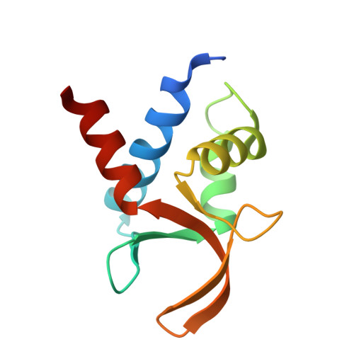
Crystal structure of TcpK in complex with oriT DNA of the antibiotic resistance plasmid pCW3.

Traore, D.A.K.Wisniewski, J.A.Flanigan, S.F.Conroy, P.J.Panjikar, S.Mok, Y.F.Lao, C.Griffin, M.D.W.Adams, V.Rood, J.I.Whisstock, J.C.

(2018) Nat Commun 9: 3732-3732

  • DOI: https://doi.org/10.1038/s41467-018-06096-2
  • Primary Citation of Related Structures:  
    5VFX, 5VFY

  • PubMed Abstract: 

    Conjugation is fundamental for the acquisition of new genetic traits and the development of antibiotic resistance in pathogenic organisms. Here, we show that a hypothetical Clostridium perfringens protein, TcpK, which is encoded by the tetracycline resistance plasmid pCW3, is essential for efficient conjugative DNA transfer. Our studies reveal that TcpK is a member of the winged helix-turn-helix (wHTH) transcription factor superfamily and that it forms a dimer in solution. Furthermore, TcpK specifically binds to a nine-nucleotide sequence that is present as tandem repeats within the pCW3 origin of transfer (oriT). The X-ray crystal structure of the TcpK-TcpK box complex reveals a binding mode centered on and around the β-wing, which is different from what has been previously shown for other wHTH proteins. Structure-guided mutagenesis experiments validate the specific interaction between TcpK and the DNA molecule. Additional studies highlight that the TcpK dimer is important for specific DNA binding.


  • Organizational Affiliation

    Department of Biochemistry and Molecular Biology, Infection and Immunity Program, Monash Biomedicine Discovery Institute, Monash University, Clayton, 3800, VIC, Australia.


Macromolecules
Find similar proteins by:  (by identity cutoff)  |  3D Structure
Entity ID: 1
MoleculeChains Sequence LengthOrganismDetailsImage
TcpK
A, B, C, D
107Clostridium perfringensMutation(s): 0 
Gene Names: ERS852446_03033pBeta2_00022pCpb2-CP1_20pCW3_0028pNetB-NE10_20pNetB_00021pTet_021
UniProt
Find proteins for Q1PLI2 (Clostridium perfringens)
Explore Q1PLI2 
Go to UniProtKB:  Q1PLI2
Entity Groups  
Sequence Clusters30% Identity50% Identity70% Identity90% Identity95% Identity100% Identity
UniProt GroupQ1PLI2
Sequence Annotations
Expand
  • Reference Sequence
Experimental Data & Validation

Experimental Data

  • Method: X-RAY DIFFRACTION
  • Resolution: 2.49 Å
  • R-Value Free: 0.233 
  • R-Value Work: 0.181 
  • R-Value Observed: 0.184 
  • Space Group: P 21 21 2
Unit Cell:
Length ( Å )Angle ( ˚ )
a = 80.56α = 90
b = 120.59β = 90
c = 52.89γ = 90
Software Package:
Software NamePurpose
Aimlessdata scaling
BUSTERrefinement
PDB_EXTRACTdata extraction
XDSdata reduction
SHELXDEphasing

Structure Validation

View Full Validation Report


Entry History 

Revision History  (Full details and data files)

  • Version 1.0: 2018-04-18
    Type: Initial release
  • Version 1.1: 2018-10-03
    Changes: Data collection, Database references
  • Version 1.2: 2024-03-13
    Changes: Data collection, Database references