5T85

Crystal structure of 10E8 Fab in complex with the MPER epitope scaffold T117v2 and phosphatidylglycerol (06:0 PG)


Experimental Data Snapshot

  • Method: X-RAY DIFFRACTION
  • Resolution: 2.37 Å
  • R-Value Free: 0.250 
  • R-Value Work: 0.195 
  • R-Value Observed: 0.198 

wwPDB Validation   3D Report Full Report


Ligand Structure Quality Assessment 


This is version 1.4 of the entry. See complete history


Literature

Lipid interactions and angle of approach to the HIV-1 viral membrane of broadly neutralizing antibody 10E8: Insights for vaccine and therapeutic design.

Irimia, A.Serra, A.M.Sarkar, A.Jacak, R.Kalyuzhniy, O.Sok, D.Saye-Francisco, K.L.Schiffner, T.Tingle, R.Kubitz, M.Adachi, Y.Stanfield, R.L.Deller, M.C.Burton, D.R.Schief, W.R.Wilson, I.A.

(2017) PLoS Pathog 13: e1006212-e1006212

  • DOI: https://doi.org/10.1371/journal.ppat.1006212
  • Primary Citation of Related Structures:  
    5SY8, 5T29, 5T5B, 5T6L, 5T80, 5T85, 5TFW

  • PubMed Abstract: 

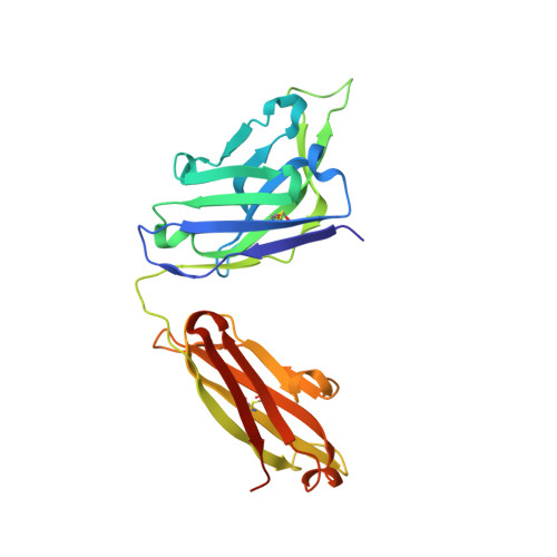
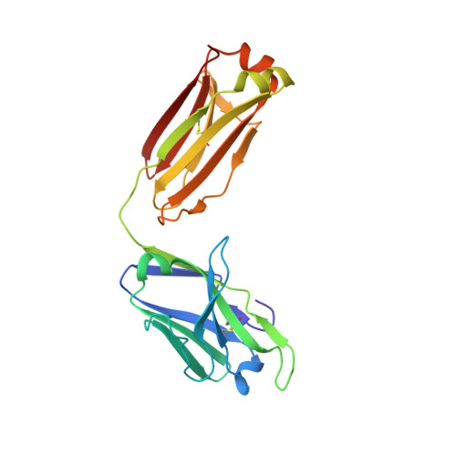
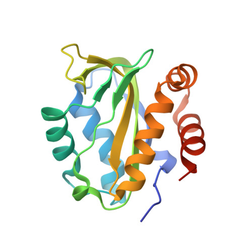
    Among broadly neutralizing antibodies to HIV, 10E8 exhibits greater neutralizing breadth than most. Consequently, this antibody is the focus of prophylactic/therapeutic development. The 10E8 epitope has been identified as the conserved membrane proximal external region (MPER) of gp41 subunit of the envelope (Env) viral glycoprotein and is a major vaccine target. However, the MPER is proximal to the viral membrane and may be laterally inserted into the membrane in the Env prefusion form. Nevertheless, 10E8 has not been reported to have significant lipid-binding reactivity. Here we report x-ray structures of lipid complexes with 10E8 and a scaffolded MPER construct and mutagenesis studies that provide evidence that the 10E8 epitope is composed of both MPER and lipid. 10E8 engages lipids through a specific lipid head group interaction site and a basic and polar surface on the light chain. In the model that we constructed, the MPER would then be essentially perpendicular to the virion membrane during 10E8 neutralization of HIV-1. As the viral membrane likely also plays a role in selecting for the germline antibody as well as size and residue composition of MPER antibody complementarity determining regions, the identification of lipid interaction sites and the MPER orientation with regard to the viral membrane surface during 10E8 engagement can be of great utility for immunogen and therapeutic design.


  • Organizational Affiliation

    Department of Integrative Structural and Computational Biology, The Scripps Research Institute, La Jolla, California, United States of America.


Macromolecules
Find similar proteins by:  (by identity cutoff)  |  3D Structure
Entity ID: 1
MoleculeChains Sequence LengthOrganismDetailsImage
Antibody 10E8 FAB HEAVY CHAINA [auth H]236Homo sapiensMutation(s): 0 
Gene Names: IGHV3-15*05
Entity Groups  
Sequence Clusters30% Identity50% Identity70% Identity90% Identity95% Identity100% Identity
Sequence Annotations
Expand
  • Reference Sequence
Find similar proteins by:  (by identity cutoff)  |  3D Structure
Entity ID: 2
MoleculeChains Sequence LengthOrganismDetailsImage
Antibody 10E8 FAB LIGHT CHAINB [auth L]215Homo sapiensMutation(s): 0 
Gene Names: IGLV3-19*01
Entity Groups  
Sequence Clusters30% Identity50% Identity70% Identity90% Identity95% Identity100% Identity
Sequence Annotations
Expand
  • Reference Sequence
Find similar proteins by:  (by identity cutoff)  |  3D Structure
Entity ID: 3
MoleculeChains Sequence LengthOrganismDetailsImage
10E8 EPITOPE SCAFFOLD T117V2C [auth G]163synthetic constructMutation(s): 0 
Entity Groups  
Sequence Clusters30% Identity50% Identity70% Identity90% Identity95% Identity100% Identity
Sequence Annotations
Expand
  • Reference Sequence
Small Molecules
Ligands 2 Unique
IDChains Name / Formula / InChI Key2D Diagram3D Interactions
44G
Query on 44G

Download Ideal Coordinates CCD File 
D [auth L](2S)-3-{[(R)-{[(2R)-2,3-dihydroxypropyl]oxy}(hydroxy)phosphoryl]oxy}-2-(hexanoyloxy)propyl hexanoate
C18 H35 O10 P
CNAHGDCINXNHER-CVEARBPZSA-N
PGE
Query on PGE

Download Ideal Coordinates CCD File 
E [auth L],
F [auth G]
TRIETHYLENE GLYCOL
C6 H14 O4
ZIBGPFATKBEMQZ-UHFFFAOYSA-N
Experimental Data & Validation

Experimental Data

  • Method: X-RAY DIFFRACTION
  • Resolution: 2.37 Å
  • R-Value Free: 0.250 
  • R-Value Work: 0.195 
  • R-Value Observed: 0.198 
  • Space Group: P 21 21 21
Unit Cell:
Length ( Å )Angle ( ˚ )
a = 55.006α = 90
b = 67.287β = 90
c = 194.678γ = 90
Software Package:
Software NamePurpose
PHENIXrefinement
HKL-2000data reduction
HKL-2000data scaling
PHASERphasing

Structure Validation

View Full Validation Report


Ligand Structure Quality Assessment 


Entry History & Funding Information

Deposition Data


Funding OrganizationLocationGrant Number
National Institutes of Health/National Institute Of Allergy and Infectious Diseases (NIH/NIAID)United StatesR01 AI084817
Scripps CHAVI-IDUnited StatesUM1 AI100663

Revision History  (Full details and data files)

  • Version 1.0: 2017-03-08
    Type: Initial release
  • Version 1.1: 2017-09-20
    Changes: Author supporting evidence
  • Version 1.2: 2017-11-01
    Changes: Author supporting evidence
  • Version 1.3: 2019-12-11
    Changes: Author supporting evidence
  • Version 1.4: 2023-10-04
    Changes: Data collection, Database references, Refinement description