5NUG

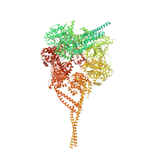
Motor domains from human cytoplasmic dynein-1 in the phi-particle conformation


Experimental Data Snapshot

  • Method: ELECTRON MICROSCOPY
  • Resolution: 3.80 Å
  • Aggregation State: PARTICLE 
  • Reconstruction Method: SINGLE PARTICLE 

wwPDB Validation   3D Report Full Report


This is version 1.3 of the entry. See complete history


Literature

Cryo-EM Reveals How Human Cytoplasmic Dynein Is Auto-inhibited and Activated.

Zhang, K.Foster, H.E.Rondelet, A.Lacey, S.E.Bahi-Buisson, N.Bird, A.W.Carter, A.P.

(2017) Cell 169: 1303-1314.e18

  • DOI: https://doi.org/10.1016/j.cell.2017.05.025
  • Primary Citation of Related Structures:  
    5NUG, 5NVS, 5NVU, 5NW4

  • PubMed Abstract: 

    Cytoplasmic dynein-1 binds dynactin and cargo adaptor proteins to form a transport machine capable of long-distance processive movement along microtubules. However, it is unclear why dynein-1 moves poorly on its own or how it is activated by dynactin. Here, we present a cryoelectron microscopy structure of the complete 1.4-megadalton human dynein-1 complex in an inhibited state known as the phi-particle. We reveal the 3D structure of the cargo binding dynein tail and show how self-dimerization of the motor domains locks them in a conformation with low microtubule affinity. Disrupting motor dimerization with structure-based mutagenesis drives dynein-1 into an open form with higher affinity for both microtubules and dynactin. We find the open form is also inhibited for movement and that dynactin relieves this by reorienting the motor domains to interact correctly with microtubules. Our model explains how dynactin binding to the dynein-1 tail directly stimulates its motor activity.


  • Organizational Affiliation

    MRC Laboratory of Molecular Biology, Cambridge CB2 0QH, UK.


Macromolecules
Find similar proteins by:  (by identity cutoff)  |  3D Structure
Entity ID: 1
MoleculeChains Sequence LengthOrganismDetailsImage
Cytoplasmic dynein 1 heavy chain 1
A, B
4,646Homo sapiensMutation(s): 0 
Gene Names: DYNC1H1DHC1DNCH1DNCLDNECLDYHCKIAA0325
UniProt & NIH Common Fund Data Resources
Find proteins for Q14204 (Homo sapiens)
Explore Q14204 
Go to UniProtKB:  Q14204
PHAROS:  Q14204
GTEx:  ENSG00000197102 
Entity Groups  
Sequence Clusters30% Identity50% Identity70% Identity90% Identity95% Identity100% Identity
UniProt GroupQ14204
Sequence Annotations
Expand
  • Reference Sequence
Small Molecules
Ligands 3 Unique
IDChains Name / Formula / InChI Key2D Diagram3D Interactions
ATP
Query on ATP

Download Ideal Coordinates CCD File 
D [auth A],
I [auth B]
ADENOSINE-5'-TRIPHOSPHATE
C10 H16 N5 O13 P3
ZKHQWZAMYRWXGA-KQYNXXCUSA-N
ADP
Query on ADP

Download Ideal Coordinates CCD File 
C [auth A]
F [auth A]
G [auth A]
H [auth B]
K [auth B]
C [auth A],
F [auth A],
G [auth A],
H [auth B],
K [auth B],
L [auth B]
ADENOSINE-5'-DIPHOSPHATE
C10 H15 N5 O10 P2
XTWYTFMLZFPYCI-KQYNXXCUSA-N
MG
Query on MG

Download Ideal Coordinates CCD File 
E [auth A],
J [auth B]
MAGNESIUM ION
Mg
JLVVSXFLKOJNIY-UHFFFAOYSA-N
Experimental Data & Validation

Experimental Data

  • Method: ELECTRON MICROSCOPY
  • Resolution: 3.80 Å
  • Aggregation State: PARTICLE 
  • Reconstruction Method: SINGLE PARTICLE 
EM Software:
TaskSoftware PackageVersion
RECONSTRUCTIONRELION1.4
MODEL REFINEMENTREFMAC5.8.0166

Structure Validation

View Full Validation Report


Entry History & Funding Information

Deposition Data


Funding OrganizationLocationGrant Number
Wellcome TrustUnited KingdomWT100387
Medical Research Council (United Kingdom)United KingdomMC_UP_A025_1011

Revision History  (Full details and data files)

  • Version 1.0: 2017-06-28
    Type: Initial release
  • Version 1.1: 2017-08-02
    Changes: Data collection, Derived calculations
  • Version 1.2: 2018-10-17
    Changes: Data collection, Refinement description
  • Version 1.3: 2019-10-23
    Changes: Data collection, Other