5NQ6

Crystal structure of the inhibited form of the redox-sensitive SufE-like sulfur acceptor CsdE from Escherichia coli at 2.40 Angstrom Resolution


Experimental Data Snapshot

  • Method: X-RAY DIFFRACTION
  • Resolution: 2.40 Å
  • R-Value Free: 0.249 
  • R-Value Work: 0.191 
  • R-Value Observed: 0.194 

wwPDB Validation   3D Report Full Report


This is version 1.0 of the entry. See complete history


Literature

Insights into the inhibited form of the redox-sensitive SufE-like sulfur acceptor CsdE.

Pena-Soler, E.Aranda, J.Lopez-Estepa, M.Gomez, S.Garces, F.Coll, M.Fernandez, F.J.Tunon, I.Vega, M.C.

(2017) PLoS One 12: e0186286-e0186286

  • DOI: https://doi.org/10.1371/journal.pone.0186286
  • Primary Citation of Related Structures:  
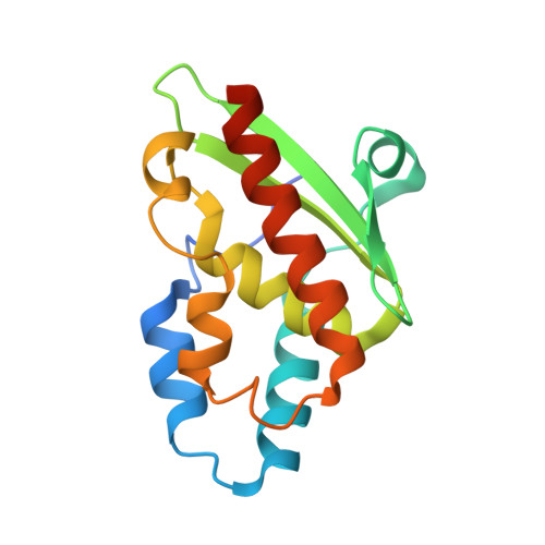
    5NQ6

  • PubMed Abstract: 

    Sulfur trafficking in living organisms relies on transpersulfuration reactions consisting in the enzyme-catalyzed transfer of S atoms via activated persulfidic S across protein-protein interfaces. The recent elucidation of the mechanistic basis for transpersulfuration in the CsdA-CsdE model system has paved the way for a better understanding of its role under oxidative stress. Herein we present the crystal structure of the oxidized, inactivated CsdE dimer at 2.4 Å resolution. The structure sheds light into the activation of the Cys61 nucleophile on its way from a solvent-secluded position in free CsdE to a fully extended conformation in the persulfurated CsdA-CsdE complex. Molecular dynamics simulations of available CsdE structures allow to delineate the sequence of conformational changes underwent by CsdE and to pinpoint the key role played by the deprotonation of the Cys61 thiol. The low-energy subunit orientation in the disulfide-bridged CsdE dimer demonstrates the likely physiologic relevance of this oxidative dead-end form of CsdE, suggesting that CsdE could act as a redox sensor in vivo.


  • Organizational Affiliation

    Chemical and Physical Biology Department, Center for Biological Research (CIB-CSIC), Madrid, Spain.


Macromolecules
Find similar proteins by:  (by identity cutoff)  |  3D Structure
Entity ID: 1
MoleculeChains Sequence LengthOrganismDetailsImage
Sulfur acceptor protein CsdE
A, B
154Escherichia coli K-12Mutation(s): 0 
Gene Names: csdEygdKb2811JW2782
UniProt
Find proteins for P0AGF2 (Escherichia coli (strain K12))
Explore P0AGF2 
Go to UniProtKB:  P0AGF2
Entity Groups  
Sequence Clusters30% Identity50% Identity70% Identity90% Identity95% Identity100% Identity
UniProt GroupP0AGF2
Sequence Annotations
Expand
  • Reference Sequence
Experimental Data & Validation

Experimental Data

  • Method: X-RAY DIFFRACTION
  • Resolution: 2.40 Å
  • R-Value Free: 0.249 
  • R-Value Work: 0.191 
  • R-Value Observed: 0.194 
  • Space Group: P 32 2 1
Unit Cell:
Length ( Å )Angle ( ˚ )
a = 58.433α = 90
b = 58.433β = 90
c = 150.452γ = 120
Software Package:
Software NamePurpose
PHENIXrefinement
XDSdata reduction
Aimlessdata scaling
AutoSolphasing

Structure Validation

View Full Validation Report


Entry History & Funding Information

Deposition Data


Funding OrganizationLocationGrant Number
Instituto de Salud Carlos IIISpainPI12/01667
Spanish Ministry of Economy and CompetitivenessSpainPET2008_0101
Spanish Ministry of Economy and CompetitivenessSpainBIO2009-11184
Spanish Ministry of Economy and CompetitivenessSpainBFU2010-22260-C02-02
Spanish Ministry of Economy and CompetitivenessSpainCTQ2015-66206-C2-2-R
Regional Government of MadridSpainS2010/BD-2316
European CommissionBelgiumComplexINC (Contract No. 279039)

Revision History  (Full details and data files)

  • Version 1.0: 2018-03-28
    Type: Initial release