5MDM

Structural intermediates in the fusion associated transition of vesiculovirus glycoprotein


Experimental Data Snapshot

  • Method: X-RAY DIFFRACTION
  • Resolution: 3.00 Å
  • R-Value Free: 0.222 
  • R-Value Work: 0.188 
  • R-Value Observed: 0.190 

wwPDB Validation   3D Report Full Report


Ligand Structure Quality Assessment 


This is version 2.1 of the entry. See complete history


Literature

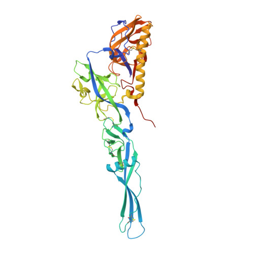
Structural intermediates in the fusion-associated transition of vesiculovirus glycoprotein.

Baquero, E.Albertini, A.A.Raux, H.Abou-Hamdan, A.Boeri-Erba, E.Ouldali, M.Buonocore, L.Rose, J.K.Lepault, J.Bressanelli, S.Gaudin, Y.

(2017) EMBO J 36: 679-692

  • DOI: https://doi.org/10.15252/embj.201694565
  • Primary Citation of Related Structures:  
    5MDM

  • PubMed Abstract: 

    Vesiculoviruses enter cells by membrane fusion, driven by a large, low-pH-induced, conformational change in the fusion glycoprotein G that involves transition from a trimeric pre-fusion toward a trimeric post-fusion state via monomeric intermediates. Here, we present the structure of the G fusion protein at intermediate pH for two vesiculoviruses, vesicular stomatitis virus (VSV) and Chandipura virus (CHAV), which is responsible for deadly encephalopathies. First, a CHAV G crystal structure shows two intermediate conformations forming a flat dimer of heterodimers. On virions, electron microscopy (EM) and tomography reveal monomeric spikes similar to one of the crystal conformations. In solution, mass spectrometry shows dimers of G. Finally, mutations at a dimer interface, involving fusion domains associated in an antiparallel manner to form an intermolecular β-sheet, affect G fusion properties. The location of the compensatory mutations restoring fusion activity strongly suggests that this interface is functionally relevant. This work reveals the range of G structural changes and suggests that G monomers can re-associate, through antiparallel interactions between fusion domains, into dimers that play a role at some early stage of the fusion process.


  • Organizational Affiliation

    Institute for Integrative Biology of the Cell (I2BC), CEA, CNRS, Univ. Paris-Sud, Université Paris-Saclay, Gif-sur-Yvette Cedex, France.


Macromolecules
Find similar proteins by:  (by identity cutoff)  |  3D Structure
Entity ID: 1
MoleculeChains Sequence LengthOrganismDetailsImage
GlycoproteinA [auth E],
B [auth F],
C [auth A],
D [auth C]
419Chandipura virusMutation(s): 0 
UniProt
Find proteins for P13180 (Chandipura virus (strain I653514))
Explore P13180 
Go to UniProtKB:  P13180
Entity Groups  
Sequence Clusters30% Identity50% Identity70% Identity90% Identity95% Identity100% Identity
UniProt GroupP13180
Sequence Annotations
Expand
  • Reference Sequence
Oligosaccharides

Help

Entity ID: 2
MoleculeChains Length2D Diagram Glycosylation3D Interactions
2-acetamido-2-deoxy-beta-D-glucopyranose-(1-4)-2-acetamido-2-deoxy-beta-D-glucopyranoseE [auth B]2N-Glycosylation
Glycosylation Resources
GlyTouCan:  G42666HT
GlyCosmos:  G42666HT
GlyGen:  G42666HT
Entity ID: 3
MoleculeChains Length2D Diagram Glycosylation3D Interactions
2-acetamido-2-deoxy-beta-D-glucopyranose-(1-4)-[alpha-L-fucopyranose-(1-6)]2-acetamido-2-deoxy-beta-D-glucopyranoseF [auth D]3N-Glycosylation
Glycosylation Resources
GlyTouCan:  G21290RB
GlyCosmos:  G21290RB
GlyGen:  G21290RB
Experimental Data & Validation

Experimental Data

  • Method: X-RAY DIFFRACTION
  • Resolution: 3.00 Å
  • R-Value Free: 0.222 
  • R-Value Work: 0.188 
  • R-Value Observed: 0.190 
  • Space Group: P 21 21 2
Unit Cell:
Length ( Å )Angle ( ˚ )
a = 150.34α = 90
b = 228.21β = 90
c = 78.84γ = 90
Software Package:
Software NamePurpose
PHENIXrefinement
XDSdata reduction
XSCALEdata scaling
PHASERphasing

Structure Validation

View Full Validation Report


Ligand Structure Quality Assessment 


Entry History & Funding Information

Deposition Data


Funding OrganizationLocationGrant Number
European UnionFranceITN VIRUS ENTRY 235649
French National Research AgencyFranceANR-08-BLAN-0256
French National Research AgencyFranceANR 11 BSV8 002 01

Revision History  (Full details and data files)

  • Version 1.0: 2016-12-14
    Type: Initial release
  • Version 1.1: 2017-02-22
    Changes: Database references
  • Version 1.2: 2017-03-15
    Changes: Database references
  • Version 1.3: 2017-08-30
    Changes: Author supporting evidence
  • Version 2.0: 2020-07-29
    Type: Remediation
    Reason: Carbohydrate remediation
    Changes: Atomic model, Data collection, Derived calculations, Structure summary
  • Version 2.1: 2024-01-17
    Changes: Data collection, Database references, Derived calculations, Refinement description, Structure summary