5JPM

Structure of the complex of human complement C4 with MASP-2 rebuilt using iMDFF


Experimental Data Snapshot

  • Method: X-RAY DIFFRACTION
  • Resolution: 3.75 Å
  • R-Value Free: 0.268 
  • R-Value Work: 0.212 
  • R-Value Observed: 0.213 

wwPDB Validation   3D Report Full Report


This is version 2.0 of the entry. See complete history

Re-refinement Note

This entry reflects an alternative modeling of the original data in: 4FXG


Literature

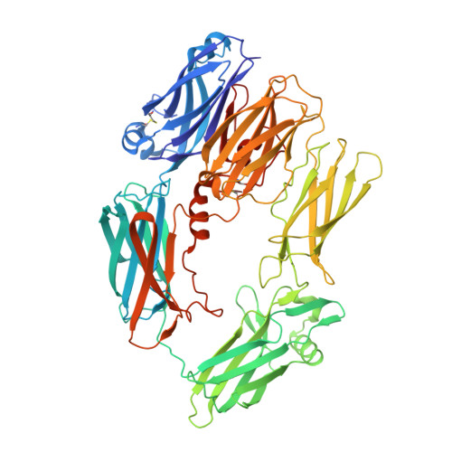
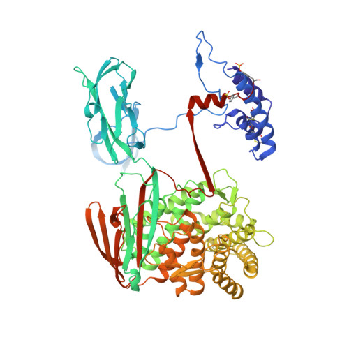
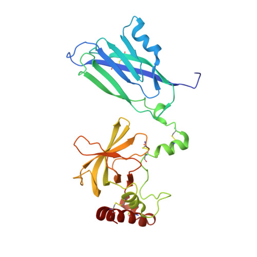
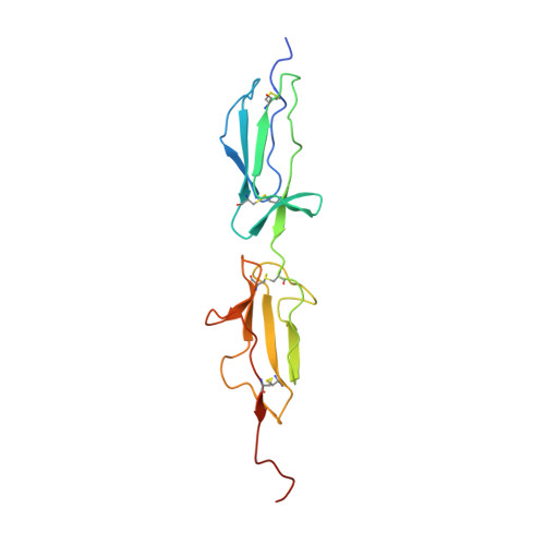
Re-evaluation of low-resolution crystal structures via interactive molecular-dynamics flexible fitting (iMDFF): a case study in complement C4.

Croll, T.I.Andersen, G.R.

(2016) Acta Crystallogr D Struct Biol 72: 1006-1016

  • DOI: https://doi.org/10.1107/S2059798316012201
  • Primary Citation of Related Structures:  
    5JPM, 5JPN, 5JTW

  • PubMed Abstract: 

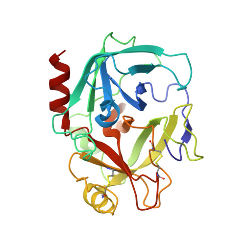
    While the rapid proliferation of high-resolution structures in the Protein Data Bank provides a rich set of templates for starting models, it remains the case that a great many structures both past and present are built at least in part by hand-threading through low-resolution and/or weak electron density. With current model-building tools this task can be challenging, and the de facto standard for acceptable error rates (in the form of atomic clashes and unfavourable backbone and side-chain conformations) in structures based on data with dmax not exceeding 3.5 Å reflects this. When combined with other factors such as model bias, these residual errors can conspire to make more serious errors in the protein fold difficult or impossible to detect. The three recently published 3.6-4.2 Å resolution structures of complement C4 (PDB entries 4fxg, 4fxk and 4xam) rank in the top quartile of structures of comparable resolution both in terms of Rfree and MolProbity score, yet, as shown here, contain register errors in six β-strands. By applying a molecular-dynamics force field that explicitly models interatomic forces and hence excludes most physically impossible conformations, the recently developed interactive molecular-dynamics flexible fitting (iMDFF) approach significantly reduces the complexity of the conformational space to be searched during manual rebuilding. This substantially improves the rate of detection and correction of register errors, and allows user-guided model building in maps with a resolution lower than 3.5 Å to converge to solutions with a stereochemical quality comparable to atomic resolution structures. Here, iMDFF has been used to individually correct and re-refine these three structures to MolProbity scores of <1.7, and strategies for working with such challenging data sets are suggested. Notably, the improved model allowed the resolution for complement C4b to be extended from 4.2 to 3.5 Å as demonstrated by paired refinement.


  • Organizational Affiliation

    Institute of Health and Biomedical Innovation, Queensland University of Technology, GPO Box 2434, Brisbane, QLD 4001, Australia.


Macromolecules
Find similar proteins by:  (by identity cutoff)  |  3D Structure
Entity ID: 1
MoleculeChains Sequence LengthOrganismDetailsImage
Complement C4-A
A, D
656Homo sapiensMutation(s): 0 
UniProt & NIH Common Fund Data Resources
Find proteins for P0C0L4 (Homo sapiens)
Explore P0C0L4 
Go to UniProtKB:  P0C0L4
PHAROS:  P0C0L4
GTEx:  ENSG00000244731 
Entity Groups  
Sequence Clusters30% Identity50% Identity70% Identity90% Identity95% Identity100% Identity
UniProt GroupP0C0L4
Sequence Annotations
Expand
  • Reference Sequence
Find similar proteins by:  (by identity cutoff)  |  3D Structure
Entity ID: 2
MoleculeChains Sequence LengthOrganismDetailsImage
Complement C4-A
B, E
767Homo sapiensMutation(s): 1 
UniProt & NIH Common Fund Data Resources
Find proteins for P0C0L4 (Homo sapiens)
Explore P0C0L4 
Go to UniProtKB:  P0C0L4
PHAROS:  P0C0L4
GTEx:  ENSG00000244731 
Entity Groups  
Sequence Clusters30% Identity50% Identity70% Identity90% Identity95% Identity100% Identity
UniProt GroupP0C0L4
Sequence Annotations
Expand
  • Reference Sequence
Find similar proteins by:  (by identity cutoff)  |  3D Structure
Entity ID: 3
MoleculeChains Sequence LengthOrganismDetailsImage
Complement C4-A
C, F
291Homo sapiensMutation(s): 0 
UniProt & NIH Common Fund Data Resources
Find proteins for P0C0L4 (Homo sapiens)
Explore P0C0L4 
Go to UniProtKB:  P0C0L4
PHAROS:  P0C0L4
GTEx:  ENSG00000244731 
Entity Groups  
Sequence Clusters30% Identity50% Identity70% Identity90% Identity95% Identity100% Identity
UniProt GroupP0C0L4
Sequence Annotations
Expand
  • Reference Sequence
Find similar proteins by:  (by identity cutoff)  |  3D Structure
Entity ID: 4
MoleculeChains Sequence LengthOrganismDetailsImage
Mannan-binding lectin serine protease 2
G, I
154Homo sapiensMutation(s): 2 
Gene Names: MASP2
EC: 3.4.21.104
UniProt & NIH Common Fund Data Resources
Find proteins for O00187 (Homo sapiens)
Explore O00187 
Go to UniProtKB:  O00187
PHAROS:  O00187
GTEx:  ENSG00000009724 
Entity Groups  
Sequence Clusters30% Identity50% Identity70% Identity90% Identity95% Identity100% Identity
UniProt GroupO00187
Sequence Annotations
Expand
  • Reference Sequence
Find similar proteins by:  (by identity cutoff)  |  3D Structure
Entity ID: 5
MoleculeChains Sequence LengthOrganismDetailsImage
Mannan-binding lectin serine protease 2
H, J
242Homo sapiensMutation(s): 1 
Gene Names: MASP2
EC: 3.4.21.104
UniProt & NIH Common Fund Data Resources
Find proteins for O00187 (Homo sapiens)
Explore O00187 
Go to UniProtKB:  O00187
PHAROS:  O00187
GTEx:  ENSG00000009724 
Entity Groups  
Sequence Clusters30% Identity50% Identity70% Identity90% Identity95% Identity100% Identity
UniProt GroupO00187
Sequence Annotations
Expand
  • Reference Sequence
Oligosaccharides

Help

Entity ID: 6
MoleculeChains Length2D Diagram Glycosylation3D Interactions
beta-D-mannopyranose-(1-4)-2-acetamido-2-deoxy-beta-D-glucopyranose-(1-4)-2-acetamido-2-deoxy-beta-D-glucopyranose
K, L, M, N, O
K, L, M, N, O, P
3N-Glycosylation
Glycosylation Resources
GlyTouCan:  G15407YE
GlyCosmos:  G15407YE
GlyGen:  G15407YE
Small Molecules
Modified Residues  1 Unique
IDChains TypeFormula2D DiagramParent
TYS
Query on TYS
B, E
L-PEPTIDE LINKINGC9 H11 N O6 STYR
Experimental Data & Validation

Experimental Data

  • Method: X-RAY DIFFRACTION
  • Resolution: 3.75 Å
  • R-Value Free: 0.268 
  • R-Value Work: 0.212 
  • R-Value Observed: 0.213 
  • Space Group: P 1 21 1
Unit Cell:
Length ( Å )Angle ( ˚ )
a = 99.01α = 90
b = 215.01β = 110.11
c = 142.86γ = 90
Software Package:
Software NamePurpose
PHENIXrefinement

Structure Validation

View Full Validation Report


Entry History 

Deposition Data

Revision History  (Full details and data files)

  • Version 1.0: 2016-08-10
    Type: Initial release
  • Version 1.1: 2016-09-14
    Changes: Database references
  • Version 1.2: 2016-09-28
    Changes: Database references
  • Version 1.3: 2019-02-20
    Changes: Advisory, Data collection, Derived calculations
  • Version 1.4: 2019-10-16
    Changes: Data collection
  • Version 2.0: 2020-07-29
    Type: Remediation
    Reason: Carbohydrate remediation
    Changes: Atomic model, Data collection, Derived calculations, Structure summary