5IK5

Laminin A2LG45 C-form, G6/7 bound.


Experimental Data Snapshot

  • Method: X-RAY DIFFRACTION
  • Resolution: 1.39 Å
  • R-Value Free: 0.177 
  • R-Value Work: 0.149 
  • R-Value Observed: 0.150 

wwPDB Validation   3D Report Full Report


Ligand Structure Quality Assessment 


This is version 2.1 of the entry. See complete history


Literature

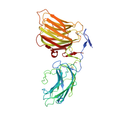
Structural basis of laminin binding to the LARGE glycans on dystroglycan.

Briggs, D.C.Yoshida-Moriguchi, T.Zheng, T.Venzke, D.Anderson, M.E.Strazzulli, A.Moracci, M.Yu, L.Hohenester, E.Campbell, K.P.

(2016) Nat Chem Biol 12: 810-814

  • DOI: https://doi.org/10.1038/nchembio.2146
  • Primary Citation of Related Structures:  
    5IK4, 5IK5, 5IK7, 5IK8

  • PubMed Abstract: 

    Dystroglycan is a highly glycosylated extracellular matrix receptor with essential functions in skeletal muscle and the nervous system. Reduced matrix binding by α-dystroglycan (α-DG) due to perturbed glycosylation is a pathological feature of several forms of muscular dystrophy. Like-acetylglucosaminyltransferase (LARGE) synthesizes the matrix-binding heteropolysaccharide [-glucuronic acid-β1,3-xylose-α1,3-]n. Using a dual exoglycosidase digestion, we confirm that this polysaccharide is present on native α-DG from skeletal muscle. The atomic details of matrix binding were revealed by a high-resolution crystal structure of laminin-G-like (LG) domains 4 and 5 (LG4 and LG5) of laminin-α2 bound to a LARGE-synthesized oligosaccharide. A single glucuronic acid-β1,3-xylose disaccharide repeat straddles a Ca(2+) ion in the LG4 domain, with oxygen atoms from both sugars replacing Ca(2+)-bound water molecules. The chelating binding mode accounts for the high affinity of this protein-carbohydrate interaction. These results reveal a previously uncharacterized mechanism of carbohydrate recognition and provide a structural framework for elucidating the mechanisms underlying muscular dystrophy.


  • Organizational Affiliation

    Department of Life Sciences, Imperial College London, London, UK.


Macromolecules
Find similar proteins by:  (by identity cutoff)  |  3D Structure
Entity ID: 1
MoleculeChains Sequence LengthOrganismDetailsImage
Laminin subunit alpha-2393Mus musculusMutation(s): 0 
Gene Names: Lama2
UniProt
Find proteins for Q60675 (Mus musculus)
Explore Q60675 
Go to UniProtKB:  Q60675
Entity Groups  
Sequence Clusters30% Identity50% Identity70% Identity90% Identity95% Identity100% Identity
UniProt GroupQ60675
Sequence Annotations
Expand
  • Reference Sequence
Oligosaccharides

Help

Entity ID: 2
MoleculeChains Length2D Diagram Glycosylation3D Interactions
beta-D-galactopyranose-(1-3)-2-acetamido-2-deoxy-alpha-D-galactopyranose
B
2O-Glycosylation
Glycosylation Resources
GlyTouCan:  G00031MO
GlyCosmos:  G00031MO
GlyGen:  G00031MO
Entity ID: 3
MoleculeChains Length2D Diagram Glycosylation3D Interactions
2-acetamido-2-deoxy-beta-D-glucopyranose-(1-4)-2-acetamido-2-deoxy-beta-D-glucopyranose
C
2N-Glycosylation
Glycosylation Resources
GlyTouCan:  G42666HT
GlyCosmos:  G42666HT
GlyGen:  G42666HT
Entity ID: 4
MoleculeChains Length2D Diagram Glycosylation3D Interactions
alpha-D-xylopyranose-(1-3)-beta-D-glucopyranuronic acid-(1-3)-alpha-D-xylopyranose-(1-3)-beta-D-glucopyranuronic acid
D
4N/A
Glycosylation Resources
GlyTouCan:  G02540QB
GlyCosmos:  G02540QB
Small Molecules
Ligands 3 Unique
IDChains Name / Formula / InChI Key2D Diagram3D Interactions
4MU
Query on 4MU

Download Ideal Coordinates CCD File 
M [auth A]7-hydroxy-4-methyl-2H-chromen-2-one
C10 H8 O3
HSHNITRMYYLLCV-UHFFFAOYSA-N
GOL
Query on GOL

Download Ideal Coordinates CCD File 
G [auth A]
H [auth A]
I [auth A]
J [auth A]
K [auth A]
G [auth A],
H [auth A],
I [auth A],
J [auth A],
K [auth A],
L [auth A]
GLYCEROL
C3 H8 O3
PEDCQBHIVMGVHV-UHFFFAOYSA-N
CA
Query on CA

Download Ideal Coordinates CCD File 
E [auth A],
F [auth A]
CALCIUM ION
Ca
BHPQYMZQTOCNFJ-UHFFFAOYSA-N
Biologically Interesting Molecules (External Reference) 1 Unique
Experimental Data & Validation

Experimental Data

  • Method: X-RAY DIFFRACTION
  • Resolution: 1.39 Å
  • R-Value Free: 0.177 
  • R-Value Work: 0.149 
  • R-Value Observed: 0.150 
  • Space Group: C 2 2 21
Unit Cell:
Length ( Å )Angle ( ˚ )
a = 70.39α = 90
b = 110.74β = 90
c = 123.9γ = 90
Software Package:
Software NamePurpose
PHENIXrefinement
XDSdata reduction
Aimlessdata scaling
PHASERphasing

Structure Validation

View Full Validation Report


Ligand Structure Quality Assessment 


Entry History & Funding Information

Deposition Data


Funding OrganizationLocationGrant Number
Wellcome TrustUnited Kingdom101748/Z/13/Z

Revision History  (Full details and data files)

  • Version 1.0: 2016-08-10
    Type: Initial release
  • Version 1.1: 2016-08-24
    Changes: Database references
  • Version 1.2: 2016-09-28
    Changes: Database references
  • Version 2.0: 2020-07-29
    Type: Remediation
    Reason: Carbohydrate remediation
    Changes: Advisory, Atomic model, Data collection, Derived calculations, Structure summary
  • Version 2.1: 2024-01-10
    Changes: Data collection, Database references, Derived calculations, Refinement description, Structure summary