5HHL

Reverse transcriptase domain of group II intron maturase from Eubacterium rectale in P21 space group


Experimental Data Snapshot

  • Method: X-RAY DIFFRACTION
  • Resolution: 2.10 Å
  • R-Value Free: 0.238 
  • R-Value Work: 0.201 
  • R-Value Observed: 0.203 

wwPDB Validation   3D Report Full Report


This is version 1.5 of the entry. See complete history


Literature

Crystal structures of a group II intron maturase reveal a missing link in spliceosome evolution.

Zhao, C.Pyle, A.M.

(2016) Nat Struct Mol Biol 23: 558-565

  • DOI: https://doi.org/10.1038/nsmb.3224
  • Primary Citation of Related Structures:  
    5HHJ, 5HHK, 5HHL, 5IRF, 5IRG

  • PubMed Abstract: 

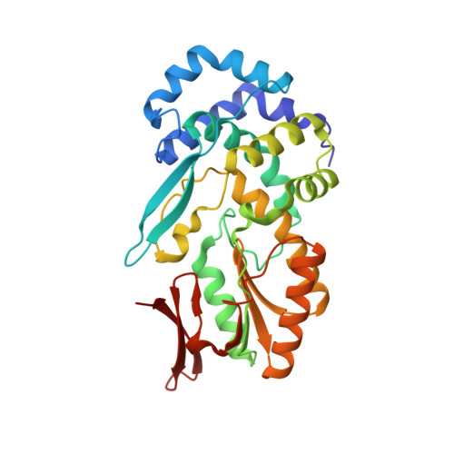
    Group II introns are self-splicing ribozymes that are essential in many organisms, and they have been hypothesized to share a common evolutionary ancestor with the spliceosome. Although structural similarity of RNA components supports this connection, it is of interest to determine whether associated protein factors also share an evolutionary heritage. Here we present the crystal structures of reverse transcriptase (RT) domains from two group II intron-encoded proteins (maturases) from Roseburia intestinalis and Eubacterium rectale, obtained at 1.2-Å and 2.1-Å resolution, respectively. These domains are more similar in architecture to the spliceosomal Prp8 RT-like domain than to any other RTs, and they share substantial similarity with flaviviral RNA polymerases. The RT domain itself is sufficient for binding intron RNA with high affinity and specificity, and it is contained within an active RT enzyme. These studies provide a foundation for understanding structure-function relationships within group II intron-maturase complexes.


  • Organizational Affiliation

    Department of Molecular Biophysics and Biochemistry, Yale University, New Haven, Connecticut, USA.


Macromolecules
Find similar proteins by:  (by identity cutoff)  |  3D Structure
Entity ID: 1
MoleculeChains Sequence LengthOrganismDetailsImage
Retron-type reverse transcriptase
A, B, C, D, E
A, B, C, D, E, F, G, H
292Agathobacter rectalis M104/1Mutation(s): 0 
Gene Names: ERE_01490
Entity Groups  
Sequence Clusters30% Identity50% Identity70% Identity90% Identity95% Identity100% Identity
Sequence Annotations
Expand
  • Reference Sequence
Experimental Data & Validation

Experimental Data

  • Method: X-RAY DIFFRACTION
  • Resolution: 2.10 Å
  • R-Value Free: 0.238 
  • R-Value Work: 0.201 
  • R-Value Observed: 0.203 
  • Space Group: P 1 21 1
Unit Cell:
Length ( Å )Angle ( ˚ )
a = 74.83α = 90
b = 119.86β = 92.08
c = 161.47γ = 90
Software Package:
Software NamePurpose
PHENIXrefinement
XDSdata reduction
XSCALEdata scaling
PHASERphasing

Structure Validation

View Full Validation Report


Entry History & Funding Information

Deposition Data


Funding OrganizationLocationGrant Number
National Institutes of Health/National Institute of General Medical Sciences (NIH/NIGMS)United StatesRO1GM50313

Revision History  (Full details and data files)

  • Version 1.0: 2016-05-04
    Type: Initial release
  • Version 1.1: 2016-05-25
    Changes: Database references
  • Version 1.2: 2016-06-22
    Changes: Database references
  • Version 1.3: 2017-09-27
    Changes: Author supporting evidence, Derived calculations
  • Version 1.4: 2019-12-25
    Changes: Author supporting evidence
  • Version 1.5: 2023-09-27
    Changes: Data collection, Database references, Derived calculations, Refinement description