The Redundancy of Peptidoglycan Carboxypeptidases Ensures Robust Cell Shape Maintenance in Escherichia Coli
Peters, K., Kannan, S., Rao, V.A., Bilboy, J., Vollmer, D., Erickson, S.W., Lewis, R.J., Young, K.D., Vollmer, W.(2016) mBio 7: 819
- PubMed: 27329754
- DOI: https://doi.org/10.1128/mBio.00819-16
- Primary Citation of Related Structures:
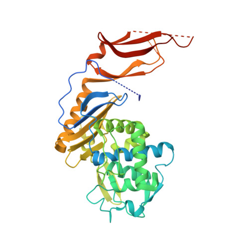
5FSR - PubMed Abstract:
Peptidoglycan (PG) is an essential structural component of the bacterial cell wall and maintains the integrity and shape of the cell by forming a continuous layer around the cytoplasmic membrane. The thin PG layer of Escherichia coli resides in the periplasm, a unique compartment whose composition and pH can vary depending on the local environment of the cell. Hence, the growth of the PG layer must be sufficiently robust to allow cell growth and division under different conditions. We have analyzed the PG composition of 28 mutants lacking multiple PG enzymes (penicillin-binding proteins [PBPs]) after growth in acidic or near-neutral-pH media. Statistical analysis of the muropeptide profiles identified dd-carboxypeptidases (DD-CPases) that were more active in cells grown at acidic pH. In particular, the absence of the DD-CPase PBP6b caused a significant increase in the pentapeptide content of PG as well as morphological defects when the cells were grown at acidic pH. Other DD-CPases (PBP4, PBP4b, PBP5, PBP6a, PBP7, and AmpH) and the PG synthase PBP1B made a smaller or null contribution to the pentapeptide-trimming activity at acidic pH. We solved the crystal structure of PBP6b and also demonstrated that the enzyme is more stable and has a lower Km at acidic pH, explaining why PBP6b is more active at low pH. Hence, PBP6b is a specialized DD-CPase that contributes to cell shape maintenance at low pH, and E. coli appears to utilize redundant DD-CPases for normal growth under different conditions.
Organizational Affiliation:
Centre for Bacterial Cell Biology, Institute for Cell and Molecular Biosciences, Newcastle University, Newcastle upon Tyne, United Kingdom.