Kinetic, Mutational, and Structural Studies of the Venezuelan Equine Encephalitis Virus Nonstructural Protein 2 Cysteine Protease.
Hu, X., Compton, J.R., Leary, D.H., Olson, M.A., Lee, M.S., Cheung, J., Ye, W., Ferrer, M., Southall, N., Jadhav, A., Morazzani, E.M., Glass, P.J., Marugan, J., Legler, P.M.(2016) Biochemistry 55: 3007-3019
- PubMed: 27030368
- DOI: https://doi.org/10.1021/acs.biochem.5b00992
- Primary Citation of Related Structures:
5EZQ, 5EZS - PubMed Abstract:
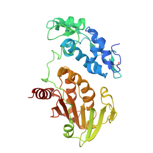
The Venezuelan equine encephalitis virus (VEEV) nonstructural protein 2 (nsP2) cysteine protease (EC 3.4.22.-) is essential for viral replication and is involved in the cytopathic effects (CPE) of the virus. The VEEV nsP2 protease is a member of MEROPS Clan CN and characteristically contains a papain-like protease linked to an S-adenosyl-l-methionine-dependent RNA methyltransferase (SAM MTase) domain. The protease contains an alternative active site motif, (475)NVCWAK(480), which differs from papain's (CGS(25)CWAFS), and the enzyme lacks a transition state-stabilizing residue homologous to Gln-19 in papain. To understand the roles of conserved residues in catalysis, we determined the structure of the free enzyme and the first structure of an inhibitor-bound alphaviral protease. The peptide-like E64d inhibitor was found to bind beneath a β-hairpin at the interface of the SAM MTase and protease domains. His-546 adopted a conformation that differed from that found in the free enzyme; one or both of the conformers may assist in leaving group departure of either the amine or Cys thiolate during the catalytic cycle. Interestingly, E64c (200 μM), the carboxylic acid form of the E64d ester, did not inhibit the nsP2 protease. To identify key residues involved in substrate binding, a number of mutants were analyzed. Mutation of the motif residue, N475A, led to a 24-fold reduction in kcat/Km, and the conformation of this residue did not change after inhibition. N475 forms a hydrogen bond with R662 in the SAM MTase domain, and the R662A and R662K mutations both led to 16-fold decreases in kcat/Km. N475 forms the base of the P1 binding site and likely orients the substrate for nucleophilic attack or plays a role in product release. An Asn homologous to N475 is similarly found in coronaviral papain-like proteases (PLpro) of the Severe Acute Respiratory Syndrome (SARS) virus and Middle East Respiratory Syndrome (MERS) virus. Mutation of another motif residue, K480A, led to a 9-fold decrease in kcat and kcat/Km. K480 likely enhances the nucleophilicity of the Cys. Consistent with our substrate-bound models, the SAM MTase domain K706A mutation increased Km 4.5-fold to 500 μM. Within the β-hairpin, the N545A mutation slightly but not significantly increased kcat and Km. The structures and identified active site residues may facilitate the discovery of protease inhibitors with antiviral activity.
Organizational Affiliation:
NIH Chemical Genomics Center, National Center for Advancing Translational Sciences , Rockville, Maryland 20850, United States.