5DET

X-ray structure of human RBPMS in complex with the RNA


Experimental Data Snapshot

  • Method: X-RAY DIFFRACTION
  • Resolution: 1.95 Å
  • R-Value Free: 0.240 
  • R-Value Work: 0.190 
  • R-Value Observed: 0.192 

wwPDB Validation   3D Report Full Report


This is version 1.3 of the entry. See complete history


Literature

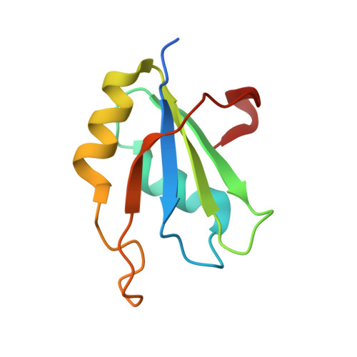
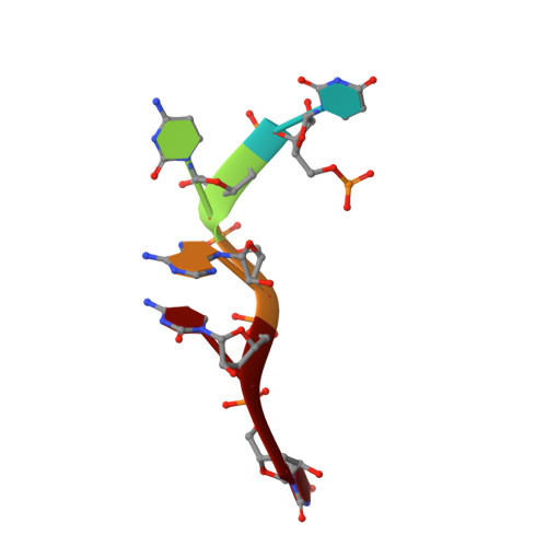
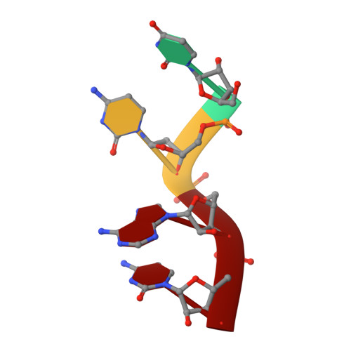
Structural basis underlying CAC RNA recognition by the RRM domain of dimeric RNA-binding protein RBPMS.

Teplova, M.Farazi, T.A.Tuschl, T.Patel, D.J.

(2016) Q Rev Biophys 49: e1-e1

  • DOI: https://doi.org/10.1017/S0033583515000207
  • Primary Citation of Related Structures:  
    5CYJ, 5DET

  • PubMed Abstract: 

    RNA-binding protein with multiple splicing (designated RBPMS) is a higher vertebrate mRNA-binding protein containing a single RNA recognition motif (RRM). RBPMS has been shown to be involved in mRNA transport, localization and stability, with key roles in axon guidance, smooth muscle plasticity, as well as regulation of cancer cell proliferation and migration. We report on structure-function studies of the RRM domain of RBPMS bound to a CAC-containing single-stranded RNA. These results provide insights into potential topologies of complexes formed by the RBPMS RRM domain and the tandem CAC repeat binding sites as detected by photoactivatable-ribonucleoside-enhanced crosslinking and immunoprecipitation. These studies establish that the RRM domain of RBPMS forms a symmetrical dimer in the free state, with each monomer binding sequence-specifically to all three nucleotides of a CAC segment in the RNA bound state. Structure-guided mutations within the dimerization and RNA-binding interfaces of RBPMS RRM on RNA complex formation resulted in both disruption of dimerization and a decrease in RNA-binding affinity as observed by size exclusion chromatography and isothermal titration calorimetry. As anticipated from biochemical binding studies, over-expression of dimerization or RNA-binding mutants of Flag-HA-tagged RBPMS were no longer able to track with stress granules in HEK293 cells, thereby documenting the deleterious effects of such mutations in vivo.


  • Organizational Affiliation

    Structural Biology Program,Memorial Sloan-Kettering Cancer Center,New York,NY 10065,USA.


Macromolecules

Find similar proteins by:  (by identity cutoff)  |  3D Structure
Entity ID: 1
MoleculeChains Sequence LengthOrganismDetailsImage
RNA-binding protein with multiple splicing
A, B
98Homo sapiensMutation(s): 0 
Gene Names: RBPMSHERMES
UniProt & NIH Common Fund Data Resources
Find proteins for Q93062 (Homo sapiens)
Explore Q93062 
Go to UniProtKB:  Q93062
PHAROS:  Q93062
GTEx:  ENSG00000157110 
Entity Groups  
Sequence Clusters30% Identity50% Identity70% Identity90% Identity95% Identity100% Identity
UniProt GroupQ93062
Sequence Annotations
Expand
  • Reference Sequence

Find similar nucleic acids by:  Sequence   |   3D Structure  

Entity ID: 2
MoleculeChains LengthOrganismImage
RNA (5'-R(P*UP*CP*AP*CP*U)-3')C [auth Q]5Homo sapiens
Sequence Annotations
Expand
  • Reference Sequence

Find similar nucleic acids by:  Sequence   |   3D Structure  

Entity ID: 3
MoleculeChains LengthOrganismImage
RNA (5'-R(*UP*CP*AP*C)-3')D [auth P]4Homo sapiens
Sequence Annotations
Expand
  • Reference Sequence
Experimental Data & Validation

Experimental Data

  • Method: X-RAY DIFFRACTION
  • Resolution: 1.95 Å
  • R-Value Free: 0.240 
  • R-Value Work: 0.190 
  • R-Value Observed: 0.192 
  • Space Group: P 1 21 1
Unit Cell:
Length ( Å )Angle ( ˚ )
a = 30.8α = 90
b = 90.232β = 93.7
c = 34.16γ = 90
Software Package:
Software NamePurpose
PHENIXrefinement
PDB_EXTRACTdata extraction
MOSFLMdata reduction
SCALAdata scaling
MOLREPphasing

Structure Validation

View Full Validation Report


Entry History 

Deposition Data

Revision History  (Full details and data files)

  • Version 1.0: 2015-09-23
    Type: Initial release
  • Version 1.1: 2016-10-05
    Changes: Database references
  • Version 1.2: 2017-09-27
    Changes: Author supporting evidence, Database references, Derived calculations
  • Version 1.3: 2023-09-27
    Changes: Data collection, Database references, Refinement description