5A8E

thermostabilised beta1-adrenoceptor with rationally designed inverse agonist 7-methylcyanopindolol bound


Experimental Data Snapshot

  • Method: X-RAY DIFFRACTION
  • Resolution: 2.40 Å
  • R-Value Free: 0.248 
  • R-Value Work: 0.217 
  • R-Value Observed: 0.218 

wwPDB Validation   3D Report Full Report


Ligand Structure Quality Assessment 


This is version 1.4 of the entry. See complete history


Literature

Pharmacological Analysis and Structure Determination of 7-Methylcyanopindolol-Bound Beta1-Adrenergic Receptor.

Sato, T.Baker, J.Warne, T.Brown, G.Leslie, A.Congreve, M.Tate, C.

(2015) Mol Pharmacol 88: 1024

  • DOI: https://doi.org/10.1124/mol.115.101030
  • Primary Citation of Related Structures:  
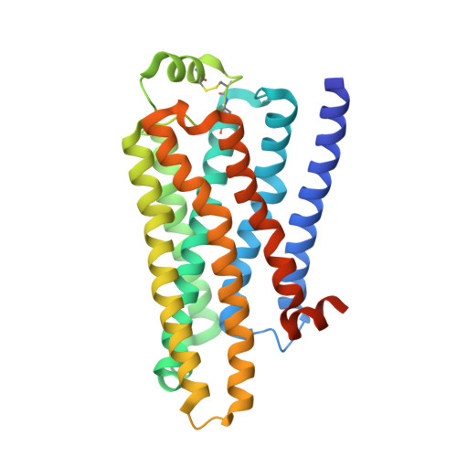
    5A8E

  • PubMed Abstract: 

    Comparisons between structures of the β1-adrenergic receptor (AR) bound to either agonists, partial agonists, or weak partial agonists led to the proposal that rotamer changes of Ser(5.46), coupled to a contraction of the binding pocket, are sufficient to increase the probability of receptor activation. (RS)-4-[3-(tert-butylamino)-2-hydroxypropoxy]-1H-indole-2-carbonitrile (cyanopindolol) is a weak partial agonist of β1AR and, based on the hypothesis above, we predicted that the addition of a methyl group to form 4-[(2S)-3-(tert-butylamino)-2-hydroxypropoxy]-7-methyl-1H-indole-2-carbonitrile (7-methylcyanopindolol) would dramatically reduce its efficacy. An eight-step synthesis of 7-methylcyanopindolol was developed and its pharmacology was analyzed. 7-Methylcyanopindolol bound with similar affinity to cyanopindolol to both β1AR and β2AR. As predicted, the efficacy of 7-methylcyanopindolol was reduced significantly compared with cyanopindolol, acting as a very weak partial agonist of turkey β1AR and an inverse agonist of human β2AR. The structure of 7-methylcyanopindolol-bound β1AR was determined to 2.4-Å resolution and found to be virtually identical to the structure of cyanopindolol-bound β1AR. The major differences in the orthosteric binding pocket are that it has expanded by 0.3 Å in 7-methylcyanopindolol-bound β1AR and the hydroxyl group of Ser(5.46) is positioned 0.8 Å further from the ligand, with respect to the position of the Ser(5.46) side chain in cyanopindolol-bound β1AR. Thus, the molecular basis for the reduction in efficacy of 7-methylcyanopindolol compared with cyanopindolol may be regarded as the opposite of the mechanism proposed for the increase in efficacy of agonists compared with antagonists.


  • Organizational Affiliation

    MRC Laboratory of Molecular Biology, Cambridge Biomedical Campus, Cambridge, United Kingdom (T.S., T.W., A.G.W.L., C.G.T.); Heptares Therapeutics Ltd, Welwyn Garden City, United Kingdom (G.A.B., M.C.); School of Life Sciences, University of Nottingham, Medical School, Queen's Medical Centre, Nottingham, United Kingdom (J.B.); KEK High Energy Accelerator Research Organization, Institute of Materials Structure Science, Structural Biology Research Center, Tsukuba, Japan (T.S.).


Macromolecules
Find similar proteins by:  (by identity cutoff)  |  3D Structure
Entity ID: 1
MoleculeChains Sequence LengthOrganismDetailsImage
BETA1 ADRENERGIC RECEPTOR315Meleagris gallopavoMutation(s): 11 
Membrane Entity: Yes 
UniProt
Find proteins for P07700 (Meleagris gallopavo)
Explore P07700 
Go to UniProtKB:  P07700
Entity Groups  
Sequence Clusters30% Identity50% Identity70% Identity90% Identity95% Identity100% Identity
UniProt GroupP07700
Sequence Annotations
Expand
  • Reference Sequence
Small Molecules
Ligands 4 Unique
IDChains Name / Formula / InChI Key2D Diagram3D Interactions
OLC
Query on OLC

Download Ideal Coordinates CCD File 
F [auth A],
G [auth A],
H [auth A],
I [auth A],
J [auth A]
(2R)-2,3-dihydroxypropyl (9Z)-octadec-9-enoate
C21 H40 O4
RZRNAYUHWVFMIP-GDCKJWNLSA-N
XTK
Query on XTK

Download Ideal Coordinates CCD File 
D [auth A]4-[(2S)-3-(tert-butylamino)-2-hydroxypropoxy]-7-methyl-1H-indole-2-carbonitrile
C17 H23 N3 O2
NDCOGBYMSDLXAU-ZDUSSCGKSA-N
MHA
Query on MHA

Download Ideal Coordinates CCD File 
E [auth A](CARBAMOYLMETHYL-CARBOXYMETHYL-AMINO)-ACETIC ACID
C6 H10 N2 O5
QZTKDVCDBIDYMD-UHFFFAOYSA-N
NA
Query on NA

Download Ideal Coordinates CCD File 
B [auth A],
C [auth A]
SODIUM ION
Na
FKNQFGJONOIPTF-UHFFFAOYSA-N
Binding Affinity Annotations 
IDSourceBinding Affinity
XTK Binding MOAD:  5A8E Kd: 0.04 (nM) from 1 assay(s)
Experimental Data & Validation

Experimental Data

  • Method: X-RAY DIFFRACTION
  • Resolution: 2.40 Å
  • R-Value Free: 0.248 
  • R-Value Work: 0.217 
  • R-Value Observed: 0.218 
  • Space Group: P 21 2 21
Unit Cell:
Length ( Å )Angle ( ˚ )
a = 53.019α = 90
b = 61.777β = 90
c = 95.561γ = 90
Software Package:
Software NamePurpose
REFMACrefinement
MOSFLMdata reduction
Aimlessdata scaling
PHASERphasing

Structure Validation

View Full Validation Report


Ligand Structure Quality Assessment 


Entry History 

Deposition Data

Revision History  (Full details and data files)

  • Version 1.0: 2015-09-30
    Type: Initial release
  • Version 1.1: 2015-11-11
    Changes: Database references
  • Version 1.2: 2019-03-06
    Changes: Data collection, Experimental preparation, Other
  • Version 1.3: 2019-04-03
    Changes: Data collection, Source and taxonomy
  • Version 1.4: 2024-01-10
    Changes: Data collection, Database references, Derived calculations, Other, Refinement description