4Z5W

The plant peptide hormone receptor


Experimental Data Snapshot

  • Method: X-RAY DIFFRACTION
  • Resolution: 2.20 Å
  • R-Value Free: 0.263 
  • R-Value Work: 0.211 
  • R-Value Observed: 0.214 

wwPDB Validation   3D Report Full Report


Ligand Structure Quality Assessment 


This is version 2.0 of the entry. See complete history


Literature

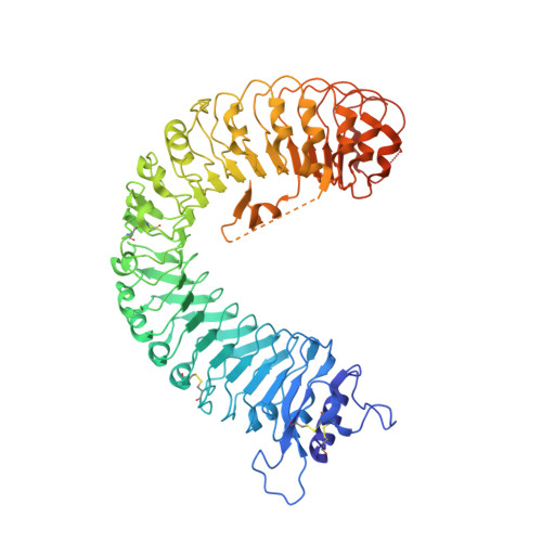
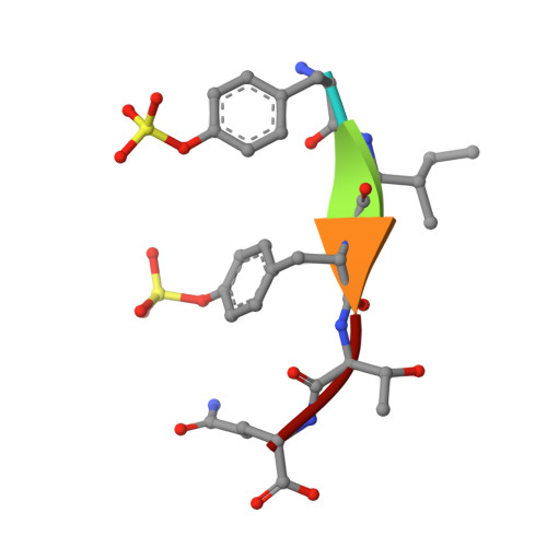
Allosteric receptor activation by the plant peptide hormone phytosulfokine

Wang, J.Li, H.Han, Z.Zhang, H.Wang, T.Lin, G.Chang, J.Yang, W.Chai, J.

(2015) Nature 525: 265-268

  • DOI: https://doi.org/10.1038/nature14858
  • Primary Citation of Related Structures:  
    4Z5W, 4Z61, 4Z62, 4Z63, 4Z64

  • PubMed Abstract: 

    Phytosulfokine (PSK) is a disulfated pentapeptide that has a ubiquitous role in plant growth and development. PSK is perceived by its receptor PSKR, a leucine-rich repeat receptor kinase (LRR-RK). The mechanisms underlying the recognition of PSK, the activation of PSKR and the identity of the components downstream of the initial binding remain elusive. Here we report the crystal structures of the extracellular LRR domain of PSKR in free, PSK- and co-receptor-bound forms. The structures reveal that PSK interacts mainly with a β-strand from the island domain of PSKR, forming an anti-β-sheet. The two sulfate moieties of PSK interact directly with PSKR, sensitizing PSKR recognition of PSK. Supported by biochemical, structural and genetic evidence, PSK binding enhances PSKR heterodimerization with the somatic embryogenesis receptor-like kinases (SERKs). However, PSK is not directly involved in PSKR-SERK interaction but stabilizes PSKR island domain for recruitment of a SERK. Our data reveal the structural basis for PSKR recognition of PSK and allosteric activation of PSKR by PSK, opening up new avenues for the design of PSKR-specific small molecules.


  • Organizational Affiliation

    Ministry of Education Key Laboratory of Protein Science, Center for Structural Biology, School of Life Sciences, Tsinghua-Peking Joint Center for Life Sciences, Tsinghua University, Beijing 100084, China.


Macromolecules
Find similar proteins by:  (by identity cutoff)  |  3D Structure
Entity ID: 1
MoleculeChains Sequence LengthOrganismDetailsImage
Phytosulfokine receptor 1
A, B
642Daucus carotaMutation(s): 0 
Gene Names: PSKR
EC: 2.7.11.1
UniProt
Find proteins for Q8LPB4 (Daucus carota)
Explore Q8LPB4 
Go to UniProtKB:  Q8LPB4
Entity Groups  
Sequence Clusters30% Identity50% Identity70% Identity90% Identity95% Identity100% Identity
UniProt GroupQ8LPB4
Sequence Annotations
Expand
  • Reference Sequence

Find similar proteins by:  Sequence   |   3D Structure  

Entity ID: 2
MoleculeChains Sequence LengthOrganismDetailsImage
PhytosulfokineC [auth P],
D [auth Q]
5ArabidopsisMutation(s): 0 
Sequence Annotations
Expand
  • Reference Sequence
Oligosaccharides

Help

Entity ID: 3
MoleculeChains Length2D Diagram Glycosylation3D Interactions
2-acetamido-2-deoxy-beta-D-glucopyranose-(1-4)-2-acetamido-2-deoxy-beta-D-glucopyranoseE [auth C]2N-Glycosylation
Glycosylation Resources
GlyTouCan:  G42666HT
GlyCosmos:  G42666HT
GlyGen:  G42666HT
Small Molecules
Ligands 1 Unique
IDChains Name / Formula / InChI Key2D Diagram3D Interactions
NAG
Query on NAG

Download Ideal Coordinates CCD File 
F [auth A]
G [auth A]
H [auth A]
I [auth A]
J [auth A]
F [auth A],
G [auth A],
H [auth A],
I [auth A],
J [auth A],
K [auth B],
L [auth B],
M [auth B],
N [auth B],
O [auth B],
P [auth B]
2-acetamido-2-deoxy-beta-D-glucopyranose
C8 H15 N O6
OVRNDRQMDRJTHS-FMDGEEDCSA-N
Modified Residues  1 Unique
IDChains TypeFormula2D DiagramParent
TYS
Query on TYS
C [auth P],
D [auth Q]
L-PEPTIDE LINKINGC9 H11 N O6 STYR
Experimental Data & Validation

Experimental Data

  • Method: X-RAY DIFFRACTION
  • Resolution: 2.20 Å
  • R-Value Free: 0.263 
  • R-Value Work: 0.211 
  • R-Value Observed: 0.214 
  • Space Group: P 1
Unit Cell:
Length ( Å )Angle ( ˚ )
a = 66.675α = 111.33
b = 75.745β = 105.71
c = 93.902γ = 97.17
Software Package:
Software NamePurpose
PHENIXrefinement
HKL-2000data scaling
PDB_EXTRACTdata extraction
HKL-2000data reduction
PHENIXphasing

Structure Validation

View Full Validation Report


Ligand Structure Quality Assessment 


Entry History 

Deposition Data

Revision History  (Full details and data files)

  • Version 1.0: 2016-03-02
    Type: Initial release
  • Version 2.0: 2020-07-29
    Type: Remediation
    Reason: Carbohydrate remediation
    Changes: Advisory, Atomic model, Data collection, Derived calculations, Structure summary