4WRL

Structure of the human CSF-1:CSF-1R complex


Experimental Data Snapshot

  • Method: X-RAY DIFFRACTION
  • Resolution: 2.80 Å
  • R-Value Free: 0.261 
  • R-Value Work: 0.223 
  • R-Value Observed: 0.225 

wwPDB Validation   3D Report Full Report


Ligand Structure Quality Assessment 


This is version 2.1 of the entry. See complete history


Literature

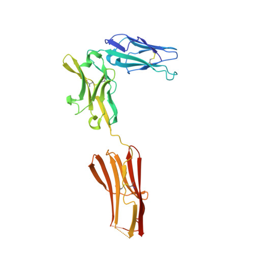
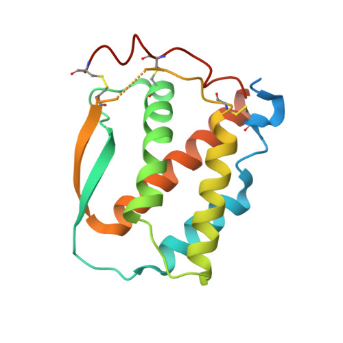
Structure and Assembly Mechanism of the Signaling Complex Mediated by Human CSF-1.

Felix, J.De Munck, S.Verstraete, K.Meuris, L.Callewaert, N.Elegheert, J.Savvides, S.N.

(2015) Structure 23: 1621-1631

  • DOI: https://doi.org/10.1016/j.str.2015.06.019
  • Primary Citation of Related Structures:  
    4WRL, 4WRM

  • PubMed Abstract: 

    Human colony-stimulating factor 1 receptor (hCSF-1R) is unique among the hematopoietic receptors because it is activated by two distinct cytokines, CSF-1 and interleukin-34 (IL-34). Despite ever-growing insights into the central role of hCSF-1R signaling in innate and adaptive immunity, inflammatory diseases, and cancer, the structural basis of the functional dichotomy of hCSF-1R has remained elusive. Here, we report crystal structures of ternary complexes between hCSF-1 and hCSF-1R, including their complete extracellular assembly, and propose a mechanism for the cooperative human CSF-1:CSF-1R complex that relies on the adoption by dimeric hCSF-1 of an active conformational state and homotypic receptor interactions. Furthermore, we trace the cytokine-binding duality of hCSF-1R to a limited set of conserved interactions mediated by functionally equivalent residues on CSF-1 and IL-34 that play into the geometric requirements of hCSF-1R activation, and map the possible mechanistic consequences of somatic mutations in hCSF-1R associated with cancer.


  • Organizational Affiliation

    Laboratory for Protein Biochemistry and Biomolecular Engineering (L-ProBE), Department of Biochemistry and Microbiology, Ghent University, 9000 Ghent, Belgium; Unit for Structural Biology, VIB Inflammation Research Center, 9052 Ghent, Belgium.


Macromolecules
Find similar proteins by:  (by identity cutoff)  |  3D Structure
Entity ID: 1
MoleculeChains Sequence LengthOrganismDetailsImage
Macrophage colony-stimulating factor 1 receptorA,
B [auth C]
286Homo sapiensMutation(s): 1 
Gene Names: CSF1RFMS
EC: 2.7.10.1
UniProt & NIH Common Fund Data Resources
Find proteins for P07333 (Homo sapiens)
Explore P07333 
Go to UniProtKB:  P07333
PHAROS:  P07333
GTEx:  ENSG00000182578 
Entity Groups  
Sequence Clusters30% Identity50% Identity70% Identity90% Identity95% Identity100% Identity
UniProt GroupP07333
Sequence Annotations
Expand
  • Reference Sequence
Find similar proteins by:  (by identity cutoff)  |  3D Structure
Entity ID: 2
MoleculeChains Sequence LengthOrganismDetailsImage
Macrophage colony-stimulating factor 1C [auth B],
D
170Homo sapiensMutation(s): 0 
Gene Names: CSF1
UniProt & NIH Common Fund Data Resources
Find proteins for P09603 (Homo sapiens)
Explore P09603 
Go to UniProtKB:  P09603
PHAROS:  P09603
GTEx:  ENSG00000184371 
Entity Groups  
Sequence Clusters30% Identity50% Identity70% Identity90% Identity95% Identity100% Identity
UniProt GroupP09603
Sequence Annotations
Expand
  • Reference Sequence
Oligosaccharides

Help

Entity ID: 3
MoleculeChains Length2D Diagram Glycosylation3D Interactions
N-acetyl-alpha-neuraminic acid-(2-3)-beta-D-galactopyranose-(1-4)-[alpha-L-fucopyranose-(1-6)]2-acetamido-2-deoxy-beta-D-glucopyranose
E, H
4N-Glycosylation
Glycosylation Resources
GlyTouCan:  G43476MN
GlyCosmos:  G43476MN
GlyGen:  G43476MN
Entity ID: 4
MoleculeChains Length2D Diagram Glycosylation3D Interactions
beta-D-galactopyranose-(1-4)-2-acetamido-2-deoxy-beta-D-glucopyranose
F
2N-Glycosylation
Glycosylation Resources
GlyTouCan:  G00055MO
GlyCosmos:  G00055MO
GlyGen:  G00055MO
Entity ID: 5
MoleculeChains Length2D Diagram Glycosylation3D Interactions
N-acetyl-alpha-neuraminic acid-(2-3)-beta-D-galactopyranose-(1-4)-2-acetamido-2-deoxy-beta-D-glucopyranose
G, I
3N-Glycosylation
Glycosylation Resources
GlyTouCan:  G00065MO
GlyCosmos:  G00065MO
GlyGen:  G00065MO
Biologically Interesting Molecules (External Reference) 1 Unique
Experimental Data & Validation

Experimental Data

  • Method: X-RAY DIFFRACTION
  • Resolution: 2.80 Å
  • R-Value Free: 0.261 
  • R-Value Work: 0.223 
  • R-Value Observed: 0.225 
  • Space Group: I 41
Unit Cell:
Length ( Å )Angle ( ˚ )
a = 142.997α = 90
b = 142.997β = 90
c = 138.323γ = 90
Software Package:
Software NamePurpose
PHENIXrefinement
XDSdata reduction
XSCALEdata scaling
PHASERphasing

Structure Validation

View Full Validation Report


Ligand Structure Quality Assessment 


Entry History 

Deposition Data

Revision History  (Full details and data files)

  • Version 1.0: 2015-08-12
    Type: Initial release
  • Version 1.1: 2015-09-09
    Changes: Database references
  • Version 2.0: 2020-07-29
    Type: Remediation
    Reason: Carbohydrate remediation
    Changes: Advisory, Atomic model, Data collection, Derived calculations, Structure summary
  • Version 2.1: 2024-01-10
    Changes: Data collection, Database references, Derived calculations, Refinement description, Structure summary