4QN9

Structure of human NAPE-PLD


Experimental Data Snapshot

  • Method: X-RAY DIFFRACTION
  • Resolution: 2.65 Å
  • R-Value Free: 0.253 
  • R-Value Work: 0.214 
  • R-Value Observed: 0.216 

wwPDB Validation   3D Report Full Report


Ligand Structure Quality Assessment 


This is version 1.2 of the entry. See complete history


Literature

Structure of human N-acylphosphatidylethanolamine-hydrolyzing phospholipase D: regulation of fatty acid ethanolamide biosynthesis by bile acids.

Magotti, P.Bauer, I.Igarashi, M.Babagoli, M.Marotta, R.Piomelli, D.Garau, G.

(2015) Structure 23: 598-604

  • DOI: https://doi.org/10.1016/j.str.2014.12.018
  • Primary Citation of Related Structures:  
    4QN9

  • PubMed Abstract: 

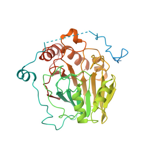
    The fatty acid ethanolamides (FAEs) are lipid mediators present in all organisms and involved in highly conserved biological functions, such as innate immunity, energy balance, and stress control. They are produced from membrane N-acylphosphatidylethanolamines (NAPEs) and include agonists for G protein-coupled receptors (e.g., cannabinoid receptors) and nuclear receptors (e.g., PPAR-α). Here, we report the crystal structure of human NAPE-hydrolyzing phospholipase D (NAPE-PLD) at 2.65 Å resolution, a membrane enzyme that catalyzes FAE formation in mammals. NAPE-PLD forms homodimers partly separated by an internal ∼ 9-Å-wide channel and uniquely adapted to associate with phospholipids. A hydrophobic cavity provides an entryway for NAPE into the active site, where a binuclear Zn(2+) center orchestrates its hydrolysis. Bile acids bind with high affinity to selective pockets in this cavity, enhancing dimer assembly and enabling catalysis. These elements offer multiple targets for the design of small-molecule NAPE-PLD modulators with potential applications in inflammation and metabolic disorders.


  • Organizational Affiliation

    Department of Drug Discovery and Development, Fondazione Istituto Italiano di Tecnologia, Via Morego 30, 16163 Genoa, Italy.


Macromolecules
Find similar proteins by:  (by identity cutoff)  |  3D Structure
Entity ID: 1
MoleculeChains Sequence LengthOrganismDetailsImage
N-acyl-phosphatidylethanolamine-hydrolyzing phospholipase D
A, B
393Homo sapiensMutation(s): 0 
Gene Names: C7orf18NAPEPLD
EC: 3.1.4.54
Membrane Entity: Yes 
UniProt & NIH Common Fund Data Resources
Find proteins for Q6IQ20 (Homo sapiens)
Explore Q6IQ20 
Go to UniProtKB:  Q6IQ20
PHAROS:  Q6IQ20
GTEx:  ENSG00000161048 
Entity Groups  
Sequence Clusters30% Identity50% Identity70% Identity90% Identity95% Identity100% Identity
UniProt GroupQ6IQ20
Sequence Annotations
Expand
  • Reference Sequence
Small Molecules
Ligands 4 Unique
IDChains Name / Formula / InChI Key2D Diagram3D Interactions
3PE
Query on 3PE

Download Ideal Coordinates CCD File 
E [auth A],
Q [auth B]
1,2-Distearoyl-sn-glycerophosphoethanolamine
C41 H82 N O8 P
LVNGJLRDBYCPGB-LDLOPFEMSA-N
DXC
Query on DXC

Download Ideal Coordinates CCD File 
F [auth A]
G [auth A]
H [auth A]
I [auth A]
L [auth B]
F [auth A],
G [auth A],
H [auth A],
I [auth A],
L [auth B],
M [auth B],
N [auth B],
R [auth B],
S [auth B],
T [auth B],
U [auth B]
(3ALPHA,5BETA,12ALPHA)-3,12-DIHYDROXYCHOLAN-24-OIC ACID
C24 H40 O4
KXGVEGMKQFWNSR-LLQZFEROSA-N
SO4
Query on SO4

Download Ideal Coordinates CCD File 
J [auth A],
K [auth A],
V [auth B]
SULFATE ION
O4 S
QAOWNCQODCNURD-UHFFFAOYSA-L
ZN
Query on ZN

Download Ideal Coordinates CCD File 
C [auth A],
D [auth A],
O [auth B],
P [auth B]
ZINC ION
Zn
PTFCDOFLOPIGGS-UHFFFAOYSA-N
Experimental Data & Validation

Experimental Data

  • Method: X-RAY DIFFRACTION
  • Resolution: 2.65 Å
  • R-Value Free: 0.253 
  • R-Value Work: 0.214 
  • R-Value Observed: 0.216 
  • Space Group: P 65 2 2
Unit Cell:
Length ( Å )Angle ( ˚ )
a = 95.099α = 90
b = 95.099β = 90
c = 444.17γ = 120
Software Package:
Software NamePurpose
CCP4refinement
MOLREPphasing
REFMACrefinement
iMOSFLMdata reduction
SCALAdata scaling

Structure Validation

View Full Validation Report


Ligand Structure Quality Assessment 


Entry History 

Deposition Data

  • Released Date: 2015-06-17 
  • Deposition Author(s): Garau, G.

Revision History  (Full details and data files)

  • Version 1.0: 2015-06-17
    Type: Initial release
  • Version 1.1: 2017-11-22
    Changes: Refinement description
  • Version 1.2: 2024-02-28
    Changes: Data collection, Database references, Derived calculations, Structure summary