Structural basis for targeting the ribosomal protein S1 of Mycobacterium tuberculosis by pyrazinamide.
Yang, J., Liu, Y., Bi, J., Cai, Q., Liao, X., Li, W., Guo, C., Zhang, Q., Lin, T., Zhao, Y., Wang, H., Liu, J., Zhang, X., Lin, D.(2015) Mol Microbiol 95: 791-803
- PubMed: 25430994
- DOI: https://doi.org/10.1111/mmi.12892
- Primary Citation of Related Structures:
4NNG, 4NNH, 4NNI, 4NNK - PubMed Abstract:
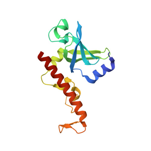
Pyrazinamide (PZA) is a first-line drug for tuberculosis (TB) treatment and is responsible for shortening the duration of TB therapy. The mode of action of PZA remains elusive. RpsA, the ribosomal protein S1 of Mycobacterium tuberculosis (Mtb), was recently identified as a target of PZA based on its binding activity to pyrazinoic acid (POA), the active form of PZA. POA binding to RpsA led to the inhibition of trans-translation. However, the nature of the RpsA-POA interaction remains unknown. Key questions include why POA exhibits an exquisite specificity to RpsA of Mtb and how RpsA mutations confer PZA resistance. Here, we report the crystal structures of the C-terminal domain of RpsA of Mtb and its complex with POA, as well as the corresponding domains of two RpsA variants that are associated with PZA resistance. Structural analysis reveals that POA binds to RpsA through hydrogen bonds and hydrophobic interactions, mediated mainly by residues (Lys303, Phe307, Phe310 and Arg357) that are essential for tmRNA binding. Conformational changes induced by mutation or sequence variation at the C-terminus of RpsA abolish the POA binding activity. Our findings provide insights into the mode of action of PZA and molecular basis of PZA resistance associated with RpsA mutations.
Organizational Affiliation:
MOE Key Laboratory of Spectrochemical Analysis & Instrumentation, Key Laboratory for Chemical Biology of Fujian Province, College of Chemistry and Chemical Engineering, Fudan University, Shanghai, 200433, China.