4MBU

Crystal structure of N-acetyltransferase from Staphylococcus aureus Mu50


Experimental Data Snapshot

  • Method: X-RAY DIFFRACTION
  • Resolution: 2.15 Å
  • R-Value Free: 0.223 
  • R-Value Work: 0.185 
  • R-Value Observed: 0.187 

wwPDB Validation   3D Report Full Report


This is version 1.4 of the entry. See complete history


Literature

Structural characterization of a Gcn5-related N-acetyltransferase from Staphylococcus aureus.

Srivastava, P.Khandokar, Y.B.Swarbrick, C.M.Roman, N.Himiari, Z.Sarker, S.Raidal, S.R.Forwood, J.K.

(2014) PLoS One 9: e102348-e102348

  • DOI: https://doi.org/10.1371/journal.pone.0102348
  • Primary Citation of Related Structures:  
    4MBU

  • PubMed Abstract: 

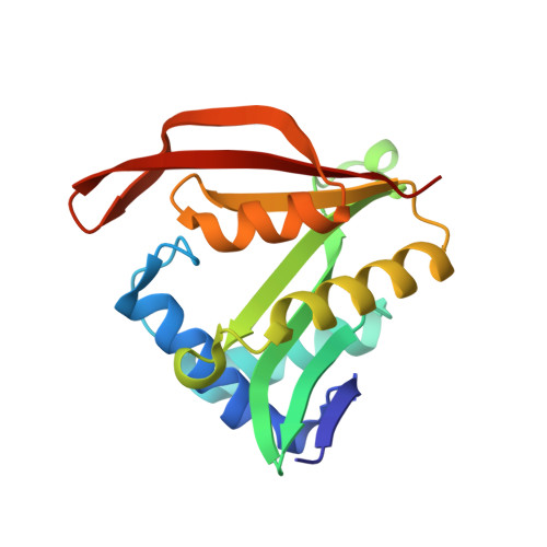
    The Gcn5-related N-acetyltransferases (GNATs) are ubiquitously expressed in nature and perform a diverse range of cellular functions through the acetylation of small molecules and protein substrates. Using activated acetyl coenzyme A as a common acetyl donor, GNATs catalyse the transfer of an acetyl group to acceptor molecules including aminoglycoside antibiotics, glucosamine-6-phosphate, histones, serotonin and spermidine. There is often only very limited sequence conservation between members of the GNAT superfamily, in part, reflecting their capacity to bind a diverse array of substrates. In contrast, the secondary and tertiary structures are highly conserved, but then at the quaternary level there is further diversity, with GNATs shown to exist in monomeric, dimeric, or tetrameric states. Here we describe the X-ray crystallographic structure of a GNAT enzyme from Staphylococcus aureus with only low sequence identity to previously solved GNAT proteins. It contains many of the classical GNAT motifs, but lacks other hallmarks of the GNAT fold including the classic β-bulge splayed at the β-sheet interface. The protein is likely to be a dimer in solution based on analysis of the asymmetric unit within the crystal structure, homology with related GNAT family members, and size exclusion chromatography. The study provides the first high resolution structure of this enzyme, providing a strong platform for substrate and cofactor modelling, and structural/functional comparisons within this diverse enzyme superfamily.


  • Organizational Affiliation

    School of Biomedical Sciences, Charles Sturt University, Wagga Wagga, New South Wales, Australia.


Macromolecules
Find similar proteins by:  (by identity cutoff)  |  3D Structure
Entity ID: 1
MoleculeChains Sequence LengthOrganismDetailsImage
Similar to N-acetyltransferase
A, B
166Staphylococcus aureus subsp. aureus Mu50Mutation(s): 0 
Gene Names: N-acetyltransferaseSAV2529
UniProt
Find proteins for A0A0H3JXG2 (Staphylococcus aureus (strain Mu50 / ATCC 700699))
Explore A0A0H3JXG2 
Go to UniProtKB:  A0A0H3JXG2
Entity Groups  
Sequence Clusters30% Identity50% Identity70% Identity90% Identity95% Identity100% Identity
UniProt GroupA0A0H3JXG2
Sequence Annotations
Expand
  • Reference Sequence
Small Molecules
Ligands 2 Unique
IDChains Name / Formula / InChI Key2D Diagram3D Interactions
CD
Query on CD

Download Ideal Coordinates CCD File 
C [auth A]
D [auth A]
E [auth A]
F [auth A]
G [auth A]
C [auth A],
D [auth A],
E [auth A],
F [auth A],
G [auth A],
I [auth B],
J [auth B],
K [auth B]
CADMIUM ION
Cd
WLZRMCYVCSSEQC-UHFFFAOYSA-N
PO4
Query on PO4

Download Ideal Coordinates CCD File 
H [auth A],
L [auth B]
PHOSPHATE ION
O4 P
NBIIXXVUZAFLBC-UHFFFAOYSA-K
Experimental Data & Validation

Experimental Data

  • Method: X-RAY DIFFRACTION
  • Resolution: 2.15 Å
  • R-Value Free: 0.223 
  • R-Value Work: 0.185 
  • R-Value Observed: 0.187 
  • Space Group: C 1 2 1
Unit Cell:
Length ( Å )Angle ( ˚ )
a = 97.49α = 90
b = 78.86β = 111.95
c = 66.01γ = 90
Software Package:
Software NamePurpose
PHENIXrefinement
CCP4model building
REFMACrefinement
MOSFLMdata reduction
SCALAdata scaling
CCP4phasing
Blu-Icedata collection

Structure Validation

View Full Validation Report


Entry History 

Deposition Data

Revision History  (Full details and data files)

  • Version 1.0: 2014-09-03
    Type: Initial release
  • Version 1.1: 2016-02-17
    Changes: Database references
  • Version 1.2: 2017-11-15
    Changes: Refinement description
  • Version 1.3: 2019-07-17
    Changes: Data collection, Refinement description
  • Version 1.4: 2024-02-28
    Changes: Data collection, Database references, Derived calculations