


Crystal structure of a nuclear GTP-binding protein from Encephalitozoon cuniculi bound to GDP-Mg2+
Edwards, T.E., Abendroth, J., Seattle Structural Genomics Center for Infectious Disease (SSGCID)To be published.
Experimental Data Snapshot
Entity ID: 1 | |||||
|---|---|---|---|---|---|
| Molecule | Chains | Sequence Length | Organism | Details | Image |
| GTP-binding nuclear protein GSP1 | 218 | Encephalitozoon cuniculi GB-M1 | Mutation(s): 0 Gene Names: ECU04_1560, GSP1 | 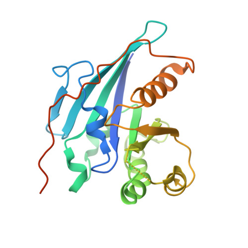 | |
UniProt | |||||
Find proteins for Q8SS11 (Encephalitozoon cuniculi (strain GB-M1)) Explore Q8SS11 Go to UniProtKB: Q8SS11 | |||||
Entity Groups | |||||
| Sequence Clusters | 30% Identity50% Identity70% Identity90% Identity95% Identity100% Identity | ||||
| UniProt Group | Q8SS11 | ||||
Sequence AnnotationsExpand | |||||
| |||||
| Ligands 3 Unique | |||||
|---|---|---|---|---|---|
| ID | Chains | Name / Formula / InChI Key | 2D Diagram | 3D Interactions | |
| GDP Query on GDP | C [auth A], E [auth B] | GUANOSINE-5'-DIPHOSPHATE C10 H15 N5 O11 P2 QGWNDRXFNXRZMB-UUOKFMHZSA-N |  | ||
| MG Query on MG | D [auth A], F [auth B] | MAGNESIUM ION Mg JLVVSXFLKOJNIY-UHFFFAOYSA-N |  | ||
| NA Query on NA | G [auth B] | SODIUM ION Na FKNQFGJONOIPTF-UHFFFAOYSA-N |  | ||
| Length ( Å ) | Angle ( ˚ ) |
|---|---|
| a = 42.57 | α = 90 |
| b = 89.25 | β = 108.06 |
| c = 54.98 | γ = 90 |
| Software Name | Purpose |
|---|---|
| XSCALE | data scaling |
| PHASER | phasing |
| REFMAC | refinement |
| PDB_EXTRACT | data extraction |
| StructureStudio | data collection |
| XDS | data reduction |
RCSB PDB (citation) is hosted by
RCSB PDB is a member of the

