Structural analysis of an oxygen-regulated diguanylate cyclase.
Tarnawski, M., Barends, T.R., Schlichting, I.(2015) Acta Crystallogr D Biol Crystallogr 71: 2158-2177
- PubMed: 26527135
- DOI: https://doi.org/10.1107/S139900471501545X
- Primary Citation of Related Structures:
4ZVA, 4ZVB, 4ZVC, 4ZVD, 4ZVE, 4ZVF, 4ZVG, 4ZVH - PubMed Abstract:
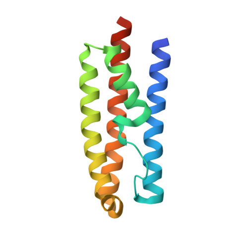
Cyclic di-GMP is a bacterial second messenger that is involved in switching between motile and sessile lifestyles. Given the medical importance of biofilm formation, there has been increasing interest in understanding the synthesis and degradation of cyclic di-GMPs and their regulation in various bacterial pathogens. Environmental cues are detected by sensing domains coupled to GGDEF and EAL or HD-GYP domains that have diguanylate cyclase and phosphodiesterase activities, respectively, producing and degrading cyclic di-GMP. The Escherichia coli protein DosC (also known as YddV) consists of an oxygen-sensing domain belonging to the class of globin sensors that is coupled to a C-terminal GGDEF domain via a previously uncharacterized middle domain. DosC is one of the most strongly expressed GGDEF proteins in E. coli, but to date structural information on this and related proteins is scarce. Here, the high-resolution structural characterization of the oxygen-sensing globin domain, the middle domain and the catalytic GGDEF domain in apo and substrate-bound forms is described. The structural changes between the iron(III) and iron(II) forms of the sensor globin domain suggest a mechanism for oxygen-dependent regulation. The structural information on the individual domains is combined into a model of the dimeric DosC holoprotein. These findings have direct implications for the oxygen-dependent regulation of the activity of the cyclase domain.
Organizational Affiliation:
Department of Biomolecular Mechanisms, Max Planck Institute for Medical Research, Jahnstrasse 29, 69120 Heidelberg, Germany.