4U8J

Structure of Aspergillus fumigatus UDP-Galactopyranose mutase mutant Y104A


Experimental Data Snapshot

  • Method: X-RAY DIFFRACTION
  • Resolution: 2.30 Å
  • R-Value Free: 0.220 
  • R-Value Work: 0.182 
  • R-Value Observed: 0.184 

wwPDB Validation   3D Report Full Report


Ligand Structure Quality Assessment 


This is version 1.4 of the entry. See complete history


Literature

Contributions of Unique Active Site Residues of Eukaryotic UDP-Galactopyranose Mutases to Substrate Recognition and Active Site Dynamics.

Da Fonseca, I.Qureshi, I.A.Mehra-Chaudhary, R.Kizjakina, K.Tanner, J.J.Sobrado, P.

(2014) Biochemistry 53: 7794-7804

  • DOI: https://doi.org/10.1021/bi501008z
  • Primary Citation of Related Structures:  
    4U8I, 4U8J, 4U8K, 4U8L, 4U8M, 4U8N, 4U8O, 4U8P, 4WX1

  • PubMed Abstract: 

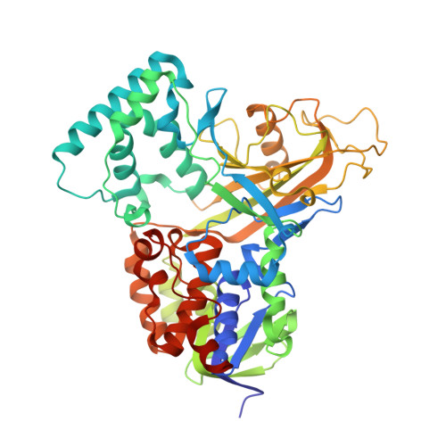
    UDP-galactopyranose mutase (UGM) catalyzes the interconversion between UDP-galactopyranose and UDP-galactofuranose. Absent in humans, galactofuranose is found in bacterial and fungal cell walls and is a cell surface virulence factor in protozoan parasites. For these reasons, UGMs are targets for drug discovery. Here, we report a mutagenesis and structural study of the UGMs from Aspergillus fumigatus and Trypanosoma cruzi focused on active site residues that are conserved in eukaryotic UGMs but are absent or different in bacterial UGMs. Kinetic analysis of the variants F66A, Y104A, Q107A, N207A, and Y317A (A. fumigatus numbering) show decreases in k(cat)/K(M) values of 200-1000-fold for the mutase reaction. In contrast, none of the mutations significantly affect the kinetics of enzyme activation by NADPH. These results indicate that the targeted residues are important for promoting the transition state conformation for UDP-galactofuranose formation. Crystal structures of the A. fumigatus mutant enzymes were determined in the presence and absence of UDP to understand the structural consequences of the mutations. The structures suggest important roles for Asn207 in stabilizing the closed active site, and Tyr317 in positioning of the uridine ring. Phe66 and the corresponding residue in Mycobacterium tuberculosis UGM (His68) play a role as the backstop, stabilizing the galactopyranose group for nucleophilic attack. Together, these results provide insight into the essentiality of the targeted residues for realizing maximal catalytic activity and a proposal for how conformational changes that close the active site are temporally related and coupled together.


  • Organizational Affiliation

    Department of Biochemistry, Virginia Tech , Blacksburg, Virginia 24061, United States.


Macromolecules
Find similar proteins by:  (by identity cutoff)  |  3D Structure
Entity ID: 1
MoleculeChains Sequence LengthOrganismDetailsImage
UDP-galactopyranose mutase
A, B, C, D
513Aspergillus fumigatusMutation(s): 4 
Gene Names: glfglfA
EC: 5.4.99.9
UniProt
Find proteins for Q4W1X2 (Aspergillus fumigatus)
Explore Q4W1X2 
Go to UniProtKB:  Q4W1X2
Entity Groups  
Sequence Clusters30% Identity50% Identity70% Identity90% Identity95% Identity100% Identity
UniProt GroupQ4W1X2
Sequence Annotations
Expand
  • Reference Sequence
Small Molecules
Ligands 3 Unique
IDChains Name / Formula / InChI Key2D Diagram3D Interactions
FDA
Query on FDA

Download Ideal Coordinates CCD File 
BA [auth C],
E [auth A],
KA [auth D],
O [auth B]
DIHYDROFLAVINE-ADENINE DINUCLEOTIDE
C27 H35 N9 O15 P2
YPZRHBJKEMOYQH-UYBVJOGSSA-N
SO4
Query on SO4

Download Ideal Coordinates CCD File 
CA [auth C]
DA [auth C]
EA [auth C]
F [auth A]
FA [auth C]
CA [auth C],
DA [auth C],
EA [auth C],
F [auth A],
FA [auth C],
G [auth A],
GA [auth C],
H [auth A],
HA [auth C],
I [auth A],
J [auth A],
K [auth A],
LA [auth D],
MA [auth D],
NA [auth D],
OA [auth D],
P [auth B],
PA [auth D],
Q [auth B],
R [auth B],
S [auth B],
T [auth B],
U [auth B]
SULFATE ION
O4 S
QAOWNCQODCNURD-UHFFFAOYSA-L
EDO
Query on EDO

Download Ideal Coordinates CCD File 
AA [auth B]
IA [auth C]
JA [auth C]
L [auth A]
M [auth A]
AA [auth B],
IA [auth C],
JA [auth C],
L [auth A],
M [auth A],
N [auth A],
QA [auth D],
RA [auth D],
V [auth B],
W [auth B],
X [auth B],
Y [auth B],
Z [auth B]
1,2-ETHANEDIOL
C2 H6 O2
LYCAIKOWRPUZTN-UHFFFAOYSA-N
Experimental Data & Validation

Experimental Data

  • Method: X-RAY DIFFRACTION
  • Resolution: 2.30 Å
  • R-Value Free: 0.220 
  • R-Value Work: 0.182 
  • R-Value Observed: 0.184 
  • Space Group: P 65 2 2
Unit Cell:
Length ( Å )Angle ( ˚ )
a = 218.419α = 90
b = 218.419β = 90
c = 322.197γ = 120
Software Package:
Software NamePurpose
XDSdata reduction
PHENIXrefinement
PDB_EXTRACTdata extraction
PHENIXrefinement

Structure Validation

View Full Validation Report


Ligand Structure Quality Assessment 


Entry History & Funding Information

Deposition Data


Funding OrganizationLocationGrant Number
National Institutes of Health/National Institute of General Medical Sciences (NIH/NIGMS)United StatesR01GM094469

Revision History  (Full details and data files)

  • Version 1.0: 2014-12-03
    Type: Initial release
  • Version 1.1: 2014-12-24
    Changes: Database references
  • Version 1.2: 2017-09-27
    Changes: Author supporting evidence, Database references, Derived calculations, Other, Source and taxonomy
  • Version 1.3: 2019-12-25
    Changes: Author supporting evidence
  • Version 1.4: 2023-09-27
    Changes: Data collection, Database references, Refinement description