4U7C

Structure of DNA polymerase kappa in complex with benzopyrene adducted DNA


Experimental Data Snapshot

  • Method: X-RAY DIFFRACTION
  • Resolution: 2.80 Å
  • R-Value Free: 0.252 
  • R-Value Work: 0.212 
  • R-Value Observed: 0.213 

wwPDB Validation   3D Report Full Report


Ligand Structure Quality Assessment 


This is version 1.6 of the entry. See complete history


Literature

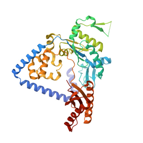
Structure and mechanism of error-free replication past the major benzo[a]pyrene adduct by human DNA polymerase kappa.

Jha, V.Bian, C.Xing, G.Ling, H.

(2016) Nucleic Acids Res 44: 4957-4967

  • DOI: https://doi.org/10.1093/nar/gkw204
  • Primary Citation of Related Structures:  
    4U6P, 4U7C

  • PubMed Abstract: 

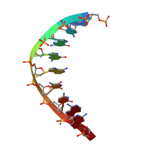
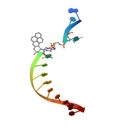
    Benzo[a]pyrene (BP) is a well-known and frequently encountered carcinogen which generates a bulky DNA adduct (+)-trans-10S-BP-N(2)-dG (BP-dG) in cells. DNA polymerase kappa (polκ) is the only known Y-family polymerase that bypasses BP-dG accurately and thus protects cells from genotoxic BP. Here, we report the structures of human polκ in complex with DNA containing either a normal guanine (G) base or a BP-dG adduct at the active site and a correct deoxycytidine. The structures and supporting biochemical data reveal a unique mechanism for accurate replication by translesion synthesis past the major bulky adduct. The active site of polκ opens at the minor groove side of the DNA substrate to accommodate the bulky BP-dG that is attached there. More importantly, polκ stabilizes the lesion DNA substrate in the same active conformation as for regular B-form DNA substrates and the bulky BPDE ring in a 5' end pointing conformation. The BP-dG adducted DNA substrate maintains a Watson-Crick (BP-dG:dC) base pair within the active site, governing correct nucleotide insertion opposite the bulky adduct. In addition, polκ's unique N-clasp domain supports the open conformation of the enzyme and the extended conformation of the single-stranded template to allow bypass of the bulky lesion. This work illustrates the first molecular mechanism for how a bulky major adduct is replicated accurately without strand misalignment and mis-insertion.


  • Organizational Affiliation

    Department of Biochemistry, Schulich School of Medicine & Dentistry, University of Western Ontario, London, Ontario, N6A 5C1, Canada.


Macromolecules

Find similar proteins by:  (by identity cutoff)  |  3D Structure
Entity ID: 1
MoleculeChains Sequence LengthOrganismDetailsImage
DNA polymerase kappa
A, B
492Homo sapiensMutation(s): 0 
Gene Names: POLKDINB1
EC: 2.7.7.7
UniProt & NIH Common Fund Data Resources
Find proteins for Q9UBT6 (Homo sapiens)
Explore Q9UBT6 
Go to UniProtKB:  Q9UBT6
PHAROS:  Q9UBT6
GTEx:  ENSG00000122008 
Entity Groups  
Sequence Clusters30% Identity50% Identity70% Identity90% Identity95% Identity100% Identity
UniProt GroupQ9UBT6
Sequence Annotations
Expand
  • Reference Sequence

Find similar nucleic acids by:  Sequence   |   3D Structure  

Entity ID: 2
MoleculeChains LengthOrganismImage
DNA (5'-D(P*GP*CP*GP*GP*AP*TP*CP*AP*G)-3')C,
E [auth P]
9unidentified
Sequence Annotations
Expand
  • Reference Sequence

Find similar nucleic acids by:  Sequence   |   3D Structure  

Entity ID: 3
MoleculeChains LengthOrganismImage
DNA (5'-D(*P*AP*TP*GP*(VKJ)P*CP*TP*GP*AP*TP*CP*CP*GP*C)-3')D,
F [auth T]
13unidentified
Sequence Annotations
Expand
  • Reference Sequence
Small Molecules
Ligands 4 Unique
IDChains Name / Formula / InChI Key2D Diagram3D Interactions
0KX
Query on 0KX

Download Ideal Coordinates CCD File 
H [auth A],
L [auth B]
2'-deoxy-5'-O-[(R)-hydroxy{[(R)-hydroxy(phosphonooxy)phosphoryl]amino}phosphoryl]cytidine
C9 H17 N4 O12 P3
STYMTWKSQLVXJN-SHYZEUOFSA-N
PEG
Query on PEG

Download Ideal Coordinates CCD File 
I [auth A],
M [auth B],
N [auth B]
DI(HYDROXYETHYL)ETHER
C4 H10 O3
MTHSVFCYNBDYFN-UHFFFAOYSA-N
EDO
Query on EDO

Download Ideal Coordinates CCD File 
J [auth B]1,2-ETHANEDIOL
C2 H6 O2
LYCAIKOWRPUZTN-UHFFFAOYSA-N
MG
Query on MG

Download Ideal Coordinates CCD File 
G [auth A],
K [auth B]
MAGNESIUM ION
Mg
JLVVSXFLKOJNIY-UHFFFAOYSA-N
Experimental Data & Validation

Experimental Data

  • Method: X-RAY DIFFRACTION
  • Resolution: 2.80 Å
  • R-Value Free: 0.252 
  • R-Value Work: 0.212 
  • R-Value Observed: 0.213 
  • Space Group: P 21 21 21
Unit Cell:
Length ( Å )Angle ( ˚ )
a = 63.64α = 90
b = 130.17β = 90
c = 166.3γ = 90
Software Package:
Software NamePurpose
PHENIXrefinement
MOSFLMdata reduction
SCALAdata scaling
PHASERphasing

Structure Validation

View Full Validation Report


Ligand Structure Quality Assessment 


Entry History & Funding Information

Deposition Data


Funding OrganizationLocationGrant Number
Canadian Institutes of Health Research (CIHR)CanadaMOP-67128

Revision History  (Full details and data files)

  • Version 1.0: 2016-01-27
    Type: Initial release
  • Version 1.1: 2016-03-23
    Changes: Derived calculations
  • Version 1.2: 2016-04-13
    Changes: Database references
  • Version 1.3: 2016-06-15
    Changes: Database references
  • Version 1.4: 2017-09-27
    Changes: Author supporting evidence, Database references, Derived calculations
  • Version 1.5: 2020-01-08
    Changes: Author supporting evidence
  • Version 1.6: 2023-09-27
    Changes: Data collection, Database references, Derived calculations, Refinement description