Molecular basis of nucleotide-dependent substrate engagement and remodeling by an AAA+ activator.
Darbari, V.C., Lawton, E., Lu, D., Burrows, P.C., Wiesler, S., Joly, N., Zhang, N., Zhang, X., Buck, M.(2014) Nucleic Acids Res 42: 9249-9261
- PubMed: 25063294
- DOI: https://doi.org/10.1093/nar/gku588
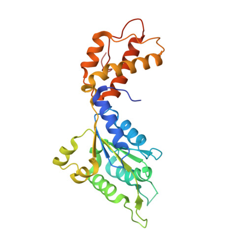
- Primary Citation of Related Structures:
4QNM, 4QNR, 4QOS - PubMed Abstract:
Binding and hydrolysis of ATP is universally required by AAA+ proteins to underpin their mechano-chemical work. Here we explore the roles of the ATPase site in an AAA+ transcriptional activator protein, the phage shock protein F (PspF), by specifically altering the Walker B motif sequence required in catalyzing ATP hydrolysis. One such mutant, the E108Q variant, is defective in ATP hydrolysis but fully remodels target transcription complexes, the RNAP-σ(54) holoenzyme, in an ATP dependent manner. Structural analysis of the E108Q variant reveals that unlike wild-type protein, which has distinct conformations for E108 residue in the ATP and ADP bound forms, E108Q adapts the same conformation irrespective of nucleotide bound. Our data show that the remodeling activities of E108Q are strongly favored on pre-melted DNA and engagement with RNAP-σ(54) using ATP binding can be sufficient to convert the inactive holoenzyme to an active form, while hydrolysis per se is required for nucleic acid remodeling that leads to transcription bubble formation. Furthermore, using linked dimer constructs, we show that RNAP-σ(54) engagement by adjacent subunits within a hexamer are required for this protein remodeling activity while DNA remodeling activity can tolerate defective ATP hydrolysis of alternating subunits.
Organizational Affiliation:
Department of Life Sciences, Imperial College London, London SW7 2AZ, UK.