4QIK

Crystal structure of the ROQ domain of human Roquin in complex with the TNF23 RNA duplex


Experimental Data Snapshot

  • Method: X-RAY DIFFRACTION
  • Resolution: 1.90 Å
  • R-Value Free: 0.212 
  • R-Value Work: 0.177 
  • R-Value Observed: 0.179 

wwPDB Validation   3D Report Full Report


This is version 1.2 of the entry. See complete history


Literature

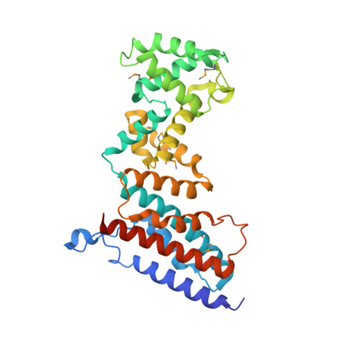
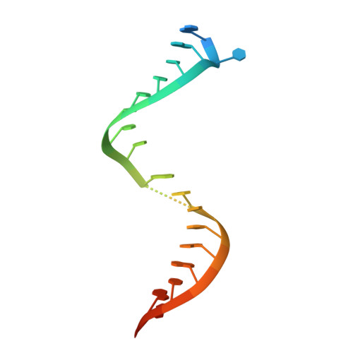
The ROQ domain of Roquin recognizes mRNA constitutive-decay element and double-stranded RNA.

Tan, D.Zhou, M.Kiledjian, M.Tong, L.

(2014) Nat Struct Mol Biol 21: 679-685

  • DOI: https://doi.org/10.1038/nsmb.2857
  • Primary Citation of Related Structures:  
    4QIK, 4QIL

  • PubMed Abstract: 

    A conserved stem-loop motif of the constitutive decay element (CDE) in the 3' UTR of mRNAs is recognized by the ROQ domain of Roquin, which mediates mRNA degradation. Here we report two crystal structures of the Homo sapiens ROQ domain in complex with CDE RNA. The ROQ domain has an elongated shape with three subdomains. The 19-nt Hmgxb3 CDE is bound as a stem-loop to domain III. The 23-nt TNF RNA is bound as a duplex to a separate site at the interface between domains I and II. Mutagenesis studies confirm that the ROQ domain has two separate RNA-binding sites, one for stem-loop RNA (A site) and the other for double-stranded RNA (B site). Mutation in either site perturbs the Roquin-mediated degradation of HMGXB3 and IL6 mRNAs in human cells, demonstrating the importance of both sites for mRNA decay.


  • Organizational Affiliation

    Department of Biological Sciences, Columbia University, New York, New York, USA.


Macromolecules

Find similar proteins by:  (by identity cutoff)  |  3D Structure
Entity ID: 1
MoleculeChains Sequence LengthOrganismDetailsImage
Roquin-1
A, B
330Homo sapiensMutation(s): 0 
Gene Names: RC3H1KIAA2025RNF198
UniProt & NIH Common Fund Data Resources
Find proteins for Q5TC82 (Homo sapiens)
Explore Q5TC82 
Go to UniProtKB:  Q5TC82
PHAROS:  Q5TC82
GTEx:  ENSG00000135870 
Entity Groups  
Sequence Clusters30% Identity50% Identity70% Identity90% Identity95% Identity100% Identity
UniProt GroupQ5TC82
Sequence Annotations
Expand
  • Reference Sequence

Find similar nucleic acids by:  Sequence   |   3D Structure  

Entity ID: 2
MoleculeChains LengthOrganismImage
5'-R(*AP*C*AP*UP*GP*UP*UP*UP*UP*CP*UP*GP*UP*GP*AP*AP*AP*AP*CP*GP*GP*AP*G)-3'
C, D
23N/A
Sequence Annotations
Expand
  • Reference Sequence
Small Molecules
Ligands 2 Unique
IDChains Name / Formula / InChI Key2D Diagram3D Interactions
GOL
Query on GOL

Download Ideal Coordinates CCD File 
S [auth B]GLYCEROL
C3 H8 O3
PEDCQBHIVMGVHV-UHFFFAOYSA-N
CL
Query on CL

Download Ideal Coordinates CCD File 
E [auth A]
F [auth A]
G [auth A]
H [auth A]
I [auth A]
E [auth A],
F [auth A],
G [auth A],
H [auth A],
I [auth A],
J [auth A],
K [auth A],
L [auth A],
M [auth B],
N [auth B],
O [auth B],
P [auth B],
Q [auth B],
R [auth B]
CHLORIDE ION
Cl
VEXZGXHMUGYJMC-UHFFFAOYSA-M
Modified Residues  1 Unique
IDChains TypeFormula2D DiagramParent
MSE
Query on MSE
A, B
L-PEPTIDE LINKINGC5 H11 N O2 SeMET
Experimental Data & Validation

Experimental Data

  • Method: X-RAY DIFFRACTION
  • Resolution: 1.90 Å
  • R-Value Free: 0.212 
  • R-Value Work: 0.177 
  • R-Value Observed: 0.179 
  • Space Group: P 21 21 21
Unit Cell:
Length ( Å )Angle ( ˚ )
a = 90.8α = 90
b = 93.1β = 90
c = 100.2γ = 90
Software Package:
Software NamePurpose
CBASSdata collection
SHELXDphasing
PHENIXrefinement
DENZOdata reduction
SCALEPACKdata scaling

Structure Validation

View Full Validation Report


Entry History 

Deposition Data

Revision History  (Full details and data files)

  • Version 1.0: 2014-07-16
    Type: Initial release
  • Version 1.1: 2014-07-30
    Changes: Database references
  • Version 1.2: 2014-08-20
    Changes: Database references