


X-ray Crystal Structure of a Putative Hydrolase from Rickettsia typhi
Fairman, J.W., Abendroth, J., Edwards, T.E., Lorimer, D.To be published.
Experimental Data Snapshot
wwPDB Validation 3D Report Full Report

Entity ID: 1 | |||||
|---|---|---|---|---|---|
| Molecule | Chains | Sequence Length | Organism | Details | Image |
| Putative Hydrolase | 298 | Rickettsia typhi str. Wilmington | Mutation(s): 0 Gene Names: RT0431 | 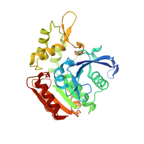 | |
UniProt | |||||
Find proteins for Q68WT4 (Rickettsia typhi (strain ATCC VR-144 / Wilmington)) Explore Q68WT4 Go to UniProtKB: Q68WT4 | |||||
Entity Groups | |||||
| Sequence Clusters | 30% Identity50% Identity70% Identity90% Identity95% Identity100% Identity | ||||
| UniProt Group | Q68WT4 | ||||
Sequence AnnotationsExpand | |||||
| |||||
| Length ( Å ) | Angle ( ˚ ) |
|---|---|
| a = 154.92 | α = 90 |
| b = 49.06 | β = 94.77 |
| c = 87.29 | γ = 90 |
| Software Name | Purpose |
|---|---|
| XSCALE | data scaling |
| PHENIX | refinement |
| PDB_EXTRACT | data extraction |
| XDS | data reduction |
| BALBES | phasing |
RCSB PDB (citation) is hosted by
RCSB PDB is a member of the

