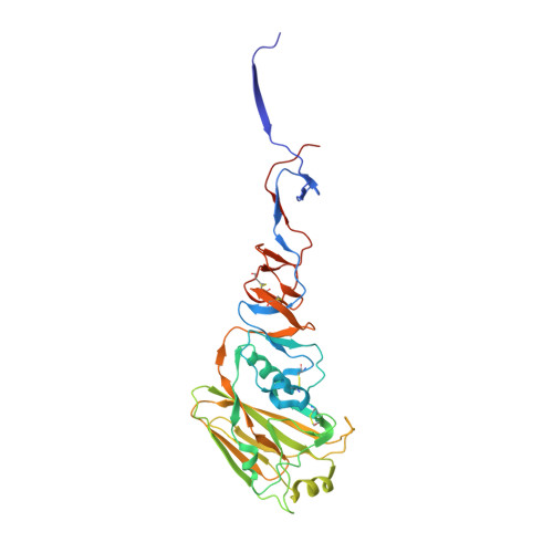
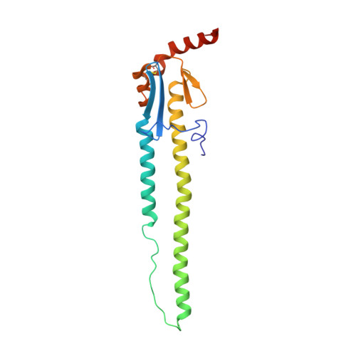
4N5Y

Crystal structure of H5 hemagglutinin mutant (N158D, N224K and Q226L) from the influenza virus A/Viet Nam/1203/2004 (H5N1)


Experimental Data Snapshot

  • Method: X-RAY DIFFRACTION
  • Resolution: 3.16 Å
  • R-Value Free: 0.262 
  • R-Value Work: 0.213 
  • R-Value Observed: 0.215 

wwPDB Validation   3D Report Full Report


Ligand Structure Quality Assessment 


This is version 2.1 of the entry. See complete history


Literature

Hemagglutinin Receptor Specificity and Structural Analyses of Respiratory Droplet-Transmissible H5N1 Viruses.

de Vries, R.P.Zhu, X.McBride, R.Rigter, A.Hanson, A.Zhong, G.Hatta, M.Xu, R.Yu, W.Kawaoka, Y.de Haan, C.A.Wilson, I.A.Paulson, J.C.

(2014) J Virol 88: 768-773

  • DOI: https://doi.org/10.1128/JVI.02690-13
  • Primary Citation of Related Structures:  
    4N5Y, 4N5Z

  • PubMed Abstract: 

    Two ferret-adapted H5N1 viruses capable of respiratory droplet transmission have been reported with mutations in the hemagglutinin receptor-binding site and stalk domains. Glycan microarray analysis reveals that both viruses exhibit a strong shift toward binding to "human-type" α2-6 sialosides but with notable differences in fine specificity. Crystal structure analysis further shows that the stalk mutation causes no obvious perturbation of the receptor-binding pocket, consistent with its impact on hemagglutinin stability without affecting receptor specificity.


  • Organizational Affiliation

    Departments of Cell and Molecular Biology and Chemical Physiology, The Scripps Research Institute, La Jolla, California, USA.


Macromolecules
Find similar proteins by:  (by identity cutoff)  |  3D Structure
Entity ID: 1
MoleculeChains Sequence LengthOrganismDetailsImage
Hemagglutinin HA1 chain334Influenza A virus (A/Viet Nam/1203/2004(H5N1))Mutation(s): 3 
Gene Names: HA
UniProt
Find proteins for Q6DQ33 (Influenza A virus (strain A/Vietnam/1203/2004 H5N1))
Explore Q6DQ33 
Go to UniProtKB:  Q6DQ33
Entity Groups  
Sequence Clusters30% Identity50% Identity70% Identity90% Identity95% Identity100% Identity
UniProt GroupQ6DQ33
Sequence Annotations
Expand
  • Reference Sequence
Find similar proteins by:  (by identity cutoff)  |  3D Structure
Entity ID: 2
MoleculeChains Sequence LengthOrganismDetailsImage
Hemagglutinin HA2 chain181Influenza A virus (A/Viet Nam/1203/2004(H5N1))Mutation(s): 0 
Gene Names: HA
UniProt
Find proteins for Q6DQ33 (Influenza A virus (strain A/Vietnam/1203/2004 H5N1))
Explore Q6DQ33 
Go to UniProtKB:  Q6DQ33
Entity Groups  
Sequence Clusters30% Identity50% Identity70% Identity90% Identity95% Identity100% Identity
UniProt GroupQ6DQ33
Sequence Annotations
Expand
  • Reference Sequence
Oligosaccharides

Help

Entity ID: 3
MoleculeChains Length2D Diagram Glycosylation3D Interactions
beta-D-mannopyranose-(1-4)-2-acetamido-2-deoxy-beta-D-glucopyranose-(1-4)-2-acetamido-2-deoxy-beta-D-glucopyranose
EA [auth e],
GA [auth g],
HA [auth h],
JA [auth j],
LA [auth l],
EA [auth e],
GA [auth g],
HA [auth h],
JA [auth j],
LA [auth l],
NA [auth n],
OA [auth o],
QA [auth q],
WA [auth w],
YA [auth y]
3N-Glycosylation
Glycosylation Resources
GlyTouCan:  G15407YE
GlyCosmos:  G15407YE
GlyGen:  G15407YE
Entity ID: 4
MoleculeChains Length2D Diagram Glycosylation3D Interactions
2-acetamido-2-deoxy-beta-D-glucopyranose-(1-4)-2-acetamido-2-deoxy-beta-D-glucopyranose
AB [auth 0],
BB [auth 1],
FA [auth f],
IA [auth i],
KA [auth k],
AB [auth 0],
BB [auth 1],
FA [auth f],
IA [auth i],
KA [auth k],
MA [auth m],
PA [auth p],
RA [auth r],
TA [auth t],
UA [auth u],
VA [auth v],
XA [auth x],
ZA [auth z]
2N-Glycosylation
Glycosylation Resources
GlyTouCan:  G42666HT
GlyCosmos:  G42666HT
GlyGen:  G42666HT
Entity ID: 5
MoleculeChains Length2D Diagram Glycosylation3D Interactions
alpha-D-mannopyranose-(1-3)-beta-D-mannopyranose-(1-4)-2-acetamido-2-deoxy-beta-D-glucopyranose-(1-4)-2-acetamido-2-deoxy-beta-D-glucopyranoseSA [auth s]4N-Glycosylation
Glycosylation Resources
GlyTouCan:  G81315DD
GlyCosmos:  G81315DD
GlyGen:  G81315DD
Experimental Data & Validation

Experimental Data

  • Method: X-RAY DIFFRACTION
  • Resolution: 3.16 Å
  • R-Value Free: 0.262 
  • R-Value Work: 0.213 
  • R-Value Observed: 0.215 
  • Space Group: P 1
Unit Cell:
Length ( Å )Angle ( ˚ )
a = 117.915α = 91.5
b = 118.105β = 90.18
c = 273.799γ = 119.87
Software Package:
Software NamePurpose
HKL-2000data collection
PHASERphasing
PHENIXrefinement
HKL-2000data reduction
HKL-2000data scaling

Structure Validation

View Full Validation Report


Ligand Structure Quality Assessment 


Entry History 

Deposition Data

Revision History  (Full details and data files)

  • Version 1.0: 2013-11-20
    Type: Initial release
  • Version 1.1: 2014-01-01
    Changes: Database references
  • Version 2.0: 2020-07-29
    Type: Remediation
    Reason: Carbohydrate remediation
    Changes: Atomic model, Data collection, Database references, Derived calculations, Structure summary
  • Version 2.1: 2023-09-20
    Changes: Data collection, Database references, Refinement description, Structure summary