Crystal structure of Ca2+/H+ antiporter protein YfkE reveals the mechanisms of Ca2+ efflux and its pH regulation.
Wu, M., Tong, S., Waltersperger, S., Diederichs, K., Wang, M., Zheng, L.(2013) Proc Natl Acad Sci U S A 110: 11367-11372
- PubMed: 23798403
- DOI: https://doi.org/10.1073/pnas.1302515110
- Primary Citation of Related Structures:
4KJR, 4KJS - PubMed Abstract:
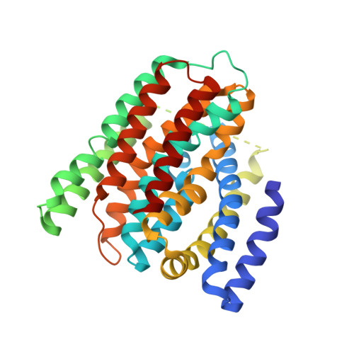
Ca(2+) efflux by Ca(2+) cation antiporter (CaCA) proteins is important for maintenance of Ca(2+) homeostasis across the cell membrane. Recently, the monomeric structure of the prokaryotic Na(+)/Ca(2+) exchanger (NCX) antiporter NCX_Mj protein from Methanococcus jannaschii shows an outward-facing conformation suggesting a hypothesis of alternating substrate access for Ca(2+) efflux. To demonstrate conformational changes essential for the CaCA mechanism, we present the crystal structure of the Ca(2+)/H(+) antiporter protein YfkE from Bacillus subtilis at 3.1-Å resolution. YfkE forms a homotrimer, confirmed by disulfide crosslinking. The protonated state of YfkE exhibits an inward-facing conformation with a large hydrophilic cavity opening to the cytoplasm in each protomer and ending in the middle of the membrane at the Ca(2+)-binding site. A hydrophobic "seal" closes its periplasmic exit. Four conserved α-repeat helices assemble in an X-like conformation to form a Ca(2+)/H(+) exchange pathway. In the Ca(2+)-binding site, two essential glutamate residues exhibit different conformations compared with their counterparts in NCX_Mj, whereas several amino acid substitutions occlude the Na(+)-binding sites. The structural differences between the inward-facing YfkE and the outward-facing NCX_Mj suggest that the conformational transition is triggered by the rotation of the kink angles of transmembrane helices 2 and 7 and is mediated by large conformational changes in their adjacent transmembrane helices 1 and 6. Our structural and mutational analyses not only establish structural bases for mechanisms of Ca(2+)/H(+) exchange and its pH regulation but also shed light on the evolutionary adaptation to different energy modes in the CaCA protein family.
Organizational Affiliation:
Center for Membrane Biology, Department of Biochemistry and Molecular Biology, University of Texas Houston Medical School, Houston, TX 77030, USA.