4K8X

Binary complex of 9N DNA polymerase in the replicative state


Experimental Data Snapshot

  • Method: X-RAY DIFFRACTION
  • Resolution: 2.28 Å
  • R-Value Free: 0.238 
  • R-Value Work: 0.195 
  • R-Value Observed: 0.198 

wwPDB Validation   3D Report Full Report


Ligand Structure Quality Assessment 


This is version 1.1 of the entry. See complete history


Literature

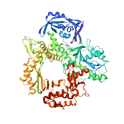
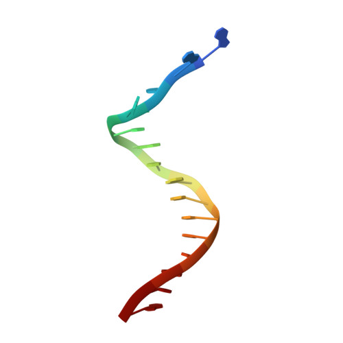
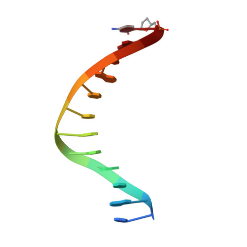
Structures of KOD and 9N DNA Polymerases Complexed with Primer Template Duplex

Bergen, K.Betz, K.Welte, W.Diederichs, K.Marx, A.

(2013) Chembiochem 14: 1058-1062

  • DOI: https://doi.org/10.1002/cbic.201300175
  • Primary Citation of Related Structures:  
    4K8X, 4K8Z

  • PubMed Abstract: 

    Replicate it: Structures of KOD and 9°N DNA polymerases, two enzymes that are widely used to replicate DNA with highly modified nucleotides, were solved at high resolution in complex with primer/template duplex. The data elucidate substrate interaction of the two enzymes and pave the way for further optimisation of the enzymes and substrates.


  • Organizational Affiliation

    Department of Chemistry, Konstanz Research School Chemical Biology, University of Konstanz, Universitätsstrasse 10, 78457 Konstanz, Germany.


Macromolecules

Find similar proteins by:  (by identity cutoff)  |  3D Structure
Entity ID: 1
MoleculeChains Sequence LengthOrganismDetailsImage
DNA polymerase775Thermococcus sp. 9oN-7Mutation(s): 2 
Gene Names: polpolA
EC: 2.7.7.7
UniProt
Find proteins for Q56366 (Thermococcus sp. (strain 9oN-7))
Explore Q56366 
Go to UniProtKB:  Q56366
Entity Groups  
Sequence Clusters30% Identity50% Identity70% Identity90% Identity95% Identity100% Identity
UniProt GroupQ56366
Sequence Annotations
Expand
  • Reference Sequence

Find similar nucleic acids by:  Sequence   |   3D Structure  

Entity ID: 2
MoleculeChains LengthOrganismImage
DNA (5'-D(P*AP*AP*AP*GP*GP*CP*GP*CP*AP*GP*TP*TP*CP*GP*CP*G)-3')B [auth T]16N/A
Sequence Annotations
Expand
  • Reference Sequence

Find similar nucleic acids by:  Sequence   |   3D Structure  

Entity ID: 3
MoleculeChains LengthOrganismImage
DNA (5'-D(*CP*GP*CP*GP*AP*AP*CP*TP*GP*CP*GP*(DOC))-3')C [auth P]12N/A
Sequence Annotations
Expand
  • Reference Sequence
Small Molecules
Ligands 4 Unique
IDChains Name / Formula / InChI Key2D Diagram3D Interactions
5CY
Query on 5CY

Download Ideal Coordinates CCD File 
P [auth T]1-(3-hydroxypropyl)-2-{(1E,3E,5E)-5-[1-(3-hydroxypropyl)-3,3-dimethyl-1,3-dihydro-2H-indol-2-ylidene]penta-1,3-dien-1-y l}-3,3-dimethyl-3H-indolium
C31 H39 N2 O2
VFBDPICHYNKCQJ-UHFFFAOYSA-N
SO4
Query on SO4

Download Ideal Coordinates CCD File 
D [auth A]
E [auth A]
F [auth A]
G [auth A]
H [auth A]
D [auth A],
E [auth A],
F [auth A],
G [auth A],
H [auth A],
I [auth A],
J [auth A],
K [auth A],
L [auth A],
N [auth A],
O [auth A]
SULFATE ION
O4 S
QAOWNCQODCNURD-UHFFFAOYSA-L
EDO
Query on EDO

Download Ideal Coordinates CCD File 
Q [auth T]1,2-ETHANEDIOL
C2 H6 O2
LYCAIKOWRPUZTN-UHFFFAOYSA-N
CL
Query on CL

Download Ideal Coordinates CCD File 
M [auth A]CHLORIDE ION
Cl
VEXZGXHMUGYJMC-UHFFFAOYSA-M
Experimental Data & Validation

Experimental Data

  • Method: X-RAY DIFFRACTION
  • Resolution: 2.28 Å
  • R-Value Free: 0.238 
  • R-Value Work: 0.195 
  • R-Value Observed: 0.198 
  • Space Group: P 21 21 2
Unit Cell:
Length ( Å )Angle ( ˚ )
a = 112.215α = 90
b = 142.616β = 90
c = 66.702γ = 90
Software Package:
Software NamePurpose
PHENIXmodel building
PHENIXrefinement
XDSdata reduction
XDSdata scaling
PHENIXphasing

Structure Validation

View Full Validation Report


Ligand Structure Quality Assessment 


Entry History 

Deposition Data

Revision History  (Full details and data files)

  • Version 1.0: 2013-06-26
    Type: Initial release
  • Version 1.1: 2023-11-08
    Changes: Data collection, Database references, Derived calculations, Refinement description, Structure summary