4JIR

Crystal Structure Of Aldose Reductase (AKR1B1) Complexed With NADP+ And Epalrestat


Experimental Data Snapshot

  • Method: X-RAY DIFFRACTION
  • Resolution: 2.00 Å
  • R-Value Free: 0.210 
  • R-Value Work: 0.160 
  • R-Value Observed: 0.160 

wwPDB Validation   3D Report Full Report


Ligand Structure Quality Assessment 


This is version 1.3 of the entry. See complete history


Literature

Inhibitor selectivity between aldo-keto reductase superfamily members AKR1B10 and AKR1B1: Role of Trp112 (Trp111).

Zhang, L.Zhang, H.Zhao, Y.Li, Z.Chen, S.Zhai, J.Chen, Y.Xie, W.Wang, Z.Li, Q.Zheng, X.Hu, X.

(2013) FEBS Lett 587: 3681-3686

  • DOI: https://doi.org/10.1016/j.febslet.2013.09.031
  • Primary Citation of Related Structures:  
    4GQG, 4I5X, 4JIH, 4JII, 4JIR

  • PubMed Abstract: 

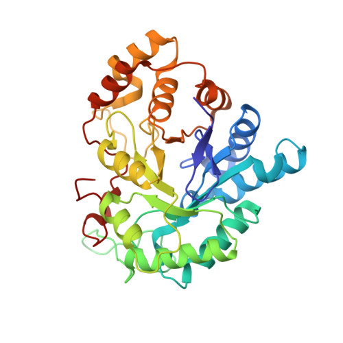
    The antineoplastic target aldo-keto reductase family member 1B10 (AKR1B10) and the critical polyol pathway enzyme aldose reductase (AKR1B1) share high structural similarity. Crystal structures reported here reveal a surprising Trp112 native conformation stabilized by a specific Gln114-centered hydrogen bond network in the AKR1B10 holoenzyme, and suggest that AKR1B1 inhibitors could retain their binding affinities toward AKR1B10 by inducing Trp112 flip to result in an "AKR1B1-like" active site in AKR1B10, while selective AKR1B10 inhibitors can take advantage of the broader active site of AKR1B10 provided by the native Trp112 side-chain orientation.


  • Organizational Affiliation

    School of Pharmaceutical Sciences, Centre for Cellular and Structural Biology of Sun Yat-sen University, Guangzhou 510006, China.


Macromolecules
Find similar proteins by:  (by identity cutoff)  |  3D Structure
Entity ID: 1
MoleculeChains Sequence LengthOrganismDetailsImage
Aldose reductase316Homo sapiensMutation(s): 0 
Gene Names: AKR1B1ALDR1
EC: 1.1.1.21
UniProt & NIH Common Fund Data Resources
Find proteins for P15121 (Homo sapiens)
Explore P15121 
Go to UniProtKB:  P15121
PHAROS:  P15121
GTEx:  ENSG00000085662 
Entity Groups  
Sequence Clusters30% Identity50% Identity70% Identity90% Identity95% Identity100% Identity
UniProt GroupP15121
Sequence Annotations
Expand
  • Reference Sequence
Small Molecules
Ligands 3 Unique
IDChains Name / Formula / InChI Key2D Diagram3D Interactions
NAP
Query on NAP

Download Ideal Coordinates CCD File 
E [auth A]NADP NICOTINAMIDE-ADENINE-DINUCLEOTIDE PHOSPHATE
C21 H28 N7 O17 P3
XJLXINKUBYWONI-NNYOXOHSSA-N
EPR
Query on EPR

Download Ideal Coordinates CCD File 
B [auth A]{5-[(2E)-2-methyl-3-phenylprop-2-en-1-ylidene]-4-oxo-2-thioxo-1,3-thiazolidin-3-yl}acetic acid
C15 H13 N O3 S2
CHNUOJQWGUIOLD-KEBJEMEDSA-N
SO4
Query on SO4

Download Ideal Coordinates CCD File 
C [auth A],
D [auth A]
SULFATE ION
O4 S
QAOWNCQODCNURD-UHFFFAOYSA-L
Binding Affinity Annotations 
IDSourceBinding Affinity
EPR Binding MOAD:  4JIR IC50: 21 (nM) from 1 assay(s)
Experimental Data & Validation

Experimental Data

  • Method: X-RAY DIFFRACTION
  • Resolution: 2.00 Å
  • R-Value Free: 0.210 
  • R-Value Work: 0.160 
  • R-Value Observed: 0.160 
  • Space Group: P 1 21 1
Unit Cell:
Length ( Å )Angle ( ˚ )
a = 47.106α = 90
b = 68.674β = 94.16
c = 49.323γ = 90
Software Package:
Software NamePurpose
CrysalisProdata collection
MOLREPphasing
PHENIXrefinement
CrysalisProdata reduction
SCALAdata scaling

Structure Validation

View Full Validation Report


Ligand Structure Quality Assessment 


Entry History 

Deposition Data

Revision History  (Full details and data files)

  • Version 1.0: 2013-10-23
    Type: Initial release
  • Version 1.1: 2013-11-27
    Changes: Database references
  • Version 1.2: 2018-01-24
    Changes: Structure summary
  • Version 1.3: 2024-02-28
    Changes: Data collection, Database references, Derived calculations