4JET

2.2A resolution structure of Holo hemophore HasA from Yersinia pestis


Experimental Data Snapshot

  • Method: X-RAY DIFFRACTION
  • Resolution: 2.20 Å
  • R-Value Free: 0.223 
  • R-Value Work: 0.166 
  • R-Value Observed: 0.169 

wwPDB Validation   3D Report Full Report


Ligand Structure Quality Assessment 


This is version 1.3 of the entry. See complete history


Literature

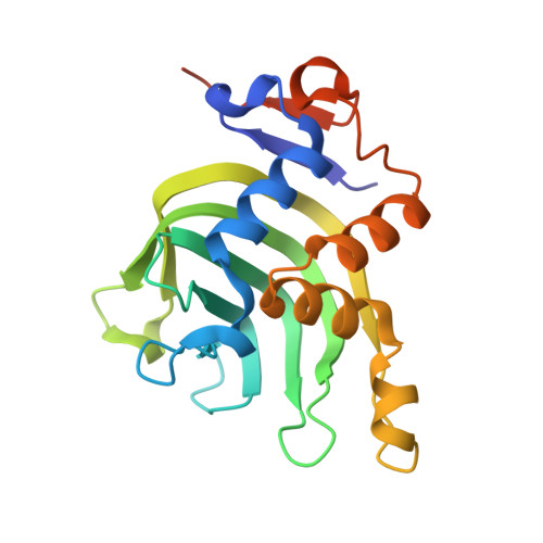
The Hemophore HasA from Yersinia pestis (HasAyp) Coordinates Hemin with a Single Residue, Tyr75, and with Minimal Conformational Change.

Kumar, R.Lovell, S.Matsumura, H.Battaile, K.P.Moenne-Loccoz, P.Rivera, M.

(2013) Biochemistry 52: 2705-2707

  • DOI: https://doi.org/10.1021/bi400280z
  • Primary Citation of Related Structures:  
    4JER, 4JES, 4JET

  • PubMed Abstract: 

    Hemophores from Serratia marcescens (HasA(sm)) and Pseudomonas aeruginosa (HasA(p)) bind hemin between two loops, which harbor the axial ligands H32 and Y75. Hemin binding to the Y75 loop triggers closing of the H32 loop and enables binding of H32. Because Yersinia pestis HasA (HasA(yp)) presents a Gln at position 32, we determined the structures of apo- and holo-HasA(yp). Surprisingly, the Q32 loop in apo-HasA(yp) is already in the closed conformation, but no residue from the Q32 loop binds hemin in holo-HasA(yp). In agreement with the minimal reorganization between the apo- and holo-structures, the hemin on-rate is too fast to detect by conventional stopped-flow measurements.


  • Organizational Affiliation

    Center for Bioinformatics, University of Kansas, 2030 Becker Drive, Lawrence, KS 66047, USA.


Macromolecules
Find similar proteins by:  (by identity cutoff)  |  3D Structure
Entity ID: 1
MoleculeChains Sequence LengthOrganismDetailsImage
Hemophore HasA
A, B, C, D, E
A, B, C, D, E, F, G, H, I, J
193Yersinia pestisMutation(s): 0 
Gene Names: hasAy0314YP_3127YPO3922
UniProt
Find proteins for Q0WA92 (Yersinia pestis)
Explore Q0WA92 
Go to UniProtKB:  Q0WA92
Entity Groups  
Sequence Clusters30% Identity50% Identity70% Identity90% Identity95% Identity100% Identity
UniProt GroupQ0WA92
Sequence Annotations
Expand
  • Reference Sequence
Small Molecules
Ligands 2 Unique
IDChains Name / Formula / InChI Key2D Diagram3D Interactions
HEM
Query on HEM

Download Ideal Coordinates CCD File 
AA [auth I]
CA [auth J]
K [auth A]
M [auth B]
O [auth C]
AA [auth I],
CA [auth J],
K [auth A],
M [auth B],
O [auth C],
Q [auth D],
S [auth E],
U [auth F],
W [auth G],
Y [auth H]
PROTOPORPHYRIN IX CONTAINING FE
C34 H32 Fe N4 O4
KABFMIBPWCXCRK-RGGAHWMASA-L
CL
Query on CL

Download Ideal Coordinates CCD File 
BA [auth I]
DA [auth J]
L [auth A]
N [auth B]
P [auth C]
BA [auth I],
DA [auth J],
L [auth A],
N [auth B],
P [auth C],
R [auth D],
T [auth E],
V [auth F],
X [auth G],
Z [auth H]
CHLORIDE ION
Cl
VEXZGXHMUGYJMC-UHFFFAOYSA-M
Experimental Data & Validation

Experimental Data

  • Method: X-RAY DIFFRACTION
  • Resolution: 2.20 Å
  • R-Value Free: 0.223 
  • R-Value Work: 0.166 
  • R-Value Observed: 0.169 
  • Space Group: C 1 2 1
Unit Cell:
Length ( Å )Angle ( ˚ )
a = 139.369α = 90
b = 69.559β = 100.12
c = 232.317γ = 90
Software Package:
Software NamePurpose
SCALAdata scaling
PHASERphasing
PHENIXrefinement
PDB_EXTRACTdata extraction
JDirectordata collection
XDSdata reduction

Structure Validation

View Full Validation Report


Ligand Structure Quality Assessment 


Entry History 

Deposition Data

Revision History  (Full details and data files)

  • Version 1.0: 2013-04-24
    Type: Initial release
  • Version 1.1: 2013-05-29
    Changes: Database references
  • Version 1.2: 2017-11-15
    Changes: Refinement description
  • Version 1.3: 2023-09-20
    Changes: Data collection, Database references, Derived calculations, Refinement description