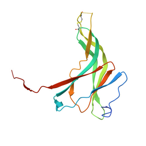
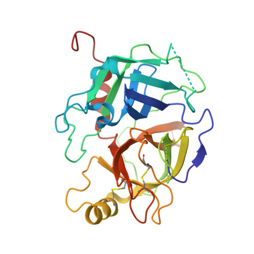
The Serine Protease Domain of MASP-3: Enzymatic Properties and Crystal Structure in Complex with Ecotin.
Gaboriaud, C., Gupta, R.K., Martin, L., Lacroix, M., Serre, L., Teillet, F., Arlaud, G.J., Rossi, V., Thielens, N.M.(2013) PLoS One 8: e67962-e67962
- PubMed: 23861840
- DOI: https://doi.org/10.1371/journal.pone.0067962
- Primary Citation of Related Structures:
4IW4 - PubMed Abstract:
Mannan-binding lectin (MBL), ficolins and collectin-11 are known to associate with three homologous modular proteases, the MBL-Associated Serine Proteases (MASPs). The crystal structures of the catalytic domains of MASP-1 and MASP-2 have been solved, but the structure of the corresponding domain of MASP-3 remains unknown. A link between mutations in the MASP1/3 gene and the rare autosomal recessive 3MC (Mingarelli, Malpuech, Michels and Carnevale,) syndrome, characterized by various developmental disorders, was discovered recently, revealing an unexpected important role of MASP-3 in early developmental processes. To gain a first insight into the enzymatic and structural properties of MASP-3, a recombinant form of its serine protease (SP) domain was produced and characterized. The amidolytic activity of this domain on fluorescent peptidyl-aminomethylcoumarin substrates was shown to be considerably lower than that of other members of the C1r/C1s/MASP family. The E. coli protease inhibitor ecotin bound to the SP domains of MASP-3 and MASP-2, whereas no significant interaction was detected with MASP-1, C1r and C1s. A tetrameric complex comprising an ecotin dimer and two MASP-3 SP domains was isolated and its crystal structure was solved and refined to 3.2 Å. Analysis of the ecotin/MASP-3 interfaces allows a better understanding of the differential reactivity of the C1r/C1s/MASP protease family members towards ecotin, and comparison of the MASP-3 SP domain structure with those of other trypsin-like proteases yields novel hypotheses accounting for its zymogen-like properties in vitro.
Organizational Affiliation:
Institut de Biologie Structurale (IBS), Direction des Sciences du Vivant, Commissariat à l'Energie Atomique et aux Energies Alternatives, Grenoble, France. christine.gaboriaud@ibs.fr