4ISZ

RNA ligase RtcB in complex with GTP alphaS and Mn(II)


Experimental Data Snapshot

  • Method: X-RAY DIFFRACTION
  • Resolution: 2.30 Å
  • R-Value Free: 0.205 
  • R-Value Work: 0.165 
  • R-Value Observed: 0.166 

wwPDB Validation   3D Report Full Report


Ligand Structure Quality Assessment 


This is version 2.1 of the entry. See complete history


Literature

Structures of the Noncanonical RNA Ligase RtcB Reveal the Mechanism of Histidine Guanylylation.

Desai, K.K.Bingman, C.A.Phillips, G.N.Raines, R.T.

(2013) Biochemistry 52: 2518-2525

  • DOI: https://doi.org/10.1021/bi4002375
  • Primary Citation of Related Structures:  
    4ISJ, 4ISZ, 4IT0

  • PubMed Abstract: 

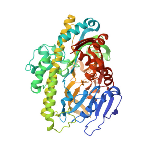
    RtcB is an atypical RNA ligase that joins either 2',3'-cyclic phosphate or 3'-phosphate termini to 5'-hydroxyl termini. In contrast to typical RNA ligases, which rely on ATP and Mg(II), catalysis by RtcB is dependent on GTP and Mn(II) with ligation proceeding through a covalent RtcB-histidine-GMP intermediate. Here, we present three structures of Pyrococcus horikoshii RtcB complexes that capture snapshots along the entire guanylylation pathway. These structures show that prior to binding GTP, a single manganese ion (Mn1) is bound to RtcB. To capture the step immediately preceding RtcB guanylylation, we determined a structure of RtcB in complex with Mn(II) and the unreactive GTP analogue guanosine 5'-(α-thio)triphosphate (GTPαS). This structure shows that Mn1 is poised to stabilize the pentavalent transition state of guanylylation while a second manganese ion (Mn2) is coordinated to a nonbridging oxygen of the γ-phosphoryl group. The pyrophosphate leaving group of GTPαS is oriented apically to His404 with the ε-nitrogen poised for in-line attack on the α-phosphorus atom. The structure of RtcB in complex with GTPαS also reveals the network of hydrogen bonds that recognize GTP and illuminates the significant conformational changes that accompany the binding of this cofactor. Finally, a structure of the enzymic histidine-GMP intermediate depicts the end of the guanylylation pathway. The ensuing molecular description of the RtcB guanylylation pathway shows that RtcB and classical ATP- and Mg(II)-dependent nucleic acid ligases have converged upon a similar two-metal mechanism for formation of the nucleotidylated enzyme intermediate.


  • Organizational Affiliation

    Department of Biochemistry, University of Wisconsin, Madison, Madison, Wisconsin 53706, USA.


Macromolecules
Find similar proteins by:  (by identity cutoff)  |  3D Structure
Entity ID: 1
MoleculeChains Sequence LengthOrganismDetailsImage
tRNA-splicing ligase RtcB
A, B
481Pyrococcus horikoshii OT3Mutation(s): 0 
Gene Names: PH1602rtcB
EC: 6.5.1 (PDB Primary Data), 3.1 (PDB Primary Data)
UniProt
Find proteins for O59245 (Pyrococcus horikoshii (strain ATCC 700860 / DSM 12428 / JCM 9974 / NBRC 100139 / OT-3))
Explore O59245 
Go to UniProtKB:  O59245
Entity Groups  
Sequence Clusters30% Identity50% Identity70% Identity90% Identity95% Identity100% Identity
UniProt GroupO59245
Sequence Annotations
Expand
  • Reference Sequence
Oligosaccharides

Help

Entity ID: 2
MoleculeChains Length2D Diagram Glycosylation3D Interactions
beta-D-fructofuranose-(2-1)-alpha-D-glucopyranose
C, D, E
2N/A
Glycosylation Resources
GlyTouCan:  G05551OP
GlyCosmos:  G05551OP
Small Molecules
Ligands 3 Unique
IDChains Name / Formula / InChI Key2D Diagram3D Interactions
GAV
Query on GAV

Download Ideal Coordinates CCD File 
F [auth A],
R [auth B]
GUANOSINE-5'-RP-ALPHA-THIO-TRIPHOSPHATE
C10 H16 N5 O13 P3 S
YRUWXBKVSXSSGS-KIOICMOXSA-N
SO4
Query on SO4

Download Ideal Coordinates CCD File 
AA [auth B]
BA [auth B]
CA [auth B]
I [auth A]
J [auth A]
AA [auth B],
BA [auth B],
CA [auth B],
I [auth A],
J [auth A],
K [auth A],
L [auth A],
M [auth A],
N [auth A],
O [auth A],
P [auth A],
Q [auth A],
U [auth B],
V [auth B],
W [auth B],
X [auth B],
Y [auth B],
Z [auth B]
SULFATE ION
O4 S
QAOWNCQODCNURD-UHFFFAOYSA-L
MN
Query on MN

Download Ideal Coordinates CCD File 
G [auth A],
H [auth A],
S [auth B],
T [auth B]
MANGANESE (II) ION
Mn
WAEMQWOKJMHJLA-UHFFFAOYSA-N
Biologically Interesting Molecules (External Reference) 1 Unique
Experimental Data & Validation

Experimental Data

  • Method: X-RAY DIFFRACTION
  • Resolution: 2.30 Å
  • R-Value Free: 0.205 
  • R-Value Work: 0.165 
  • R-Value Observed: 0.166 
  • Space Group: P 21 21 21
Unit Cell:
Length ( Å )Angle ( ˚ )
a = 81.108α = 90
b = 138.912β = 90
c = 149.469γ = 90
Software Package:
Software NamePurpose
MAR345dtbdata collection
PHENIXmodel building
PHENIXrefinement
HKL-2000data reduction
HKL-2000data scaling
PHENIXphasing

Structure Validation

View Full Validation Report


Ligand Structure Quality Assessment 


Entry History 

Deposition Data

Revision History  (Full details and data files)

  • Version 1.0: 2013-04-10
    Type: Initial release
  • Version 1.1: 2013-05-01
    Changes: Database references
  • Version 1.2: 2017-08-09
    Changes: Source and taxonomy
  • Version 2.0: 2020-07-29
    Type: Remediation
    Reason: Carbohydrate remediation
    Changes: Atomic model, Data collection, Derived calculations, Non-polymer description, Structure summary
  • Version 2.1: 2024-02-28
    Changes: Data collection, Database references, Structure summary