4HXJ

Crystal structure of SH3:RGT complex


Experimental Data Snapshot

  • Method: X-RAY DIFFRACTION
  • Resolution: 2.00 Å
  • R-Value Free: 0.193 
  • R-Value Work: 0.139 
  • R-Value Observed: 0.144 

wwPDB Validation   3D Report Full Report


This is version 1.3 of the entry. See complete history


Literature

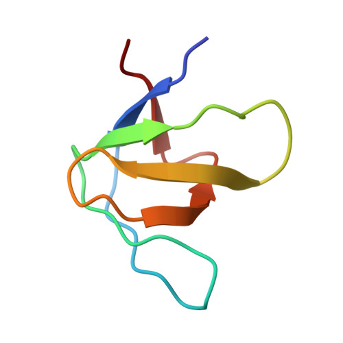
Structural framework of c-Src activation by integrin beta 3

Xiao, R.Xi, X.D.Chen, Z.Chen, S.J.Meng, G.

(2013) Blood 121: 700-706

  • DOI: https://doi.org/10.1182/blood-2012-07-440644
  • Primary Citation of Related Structures:  
    4HXJ

  • PubMed Abstract: 

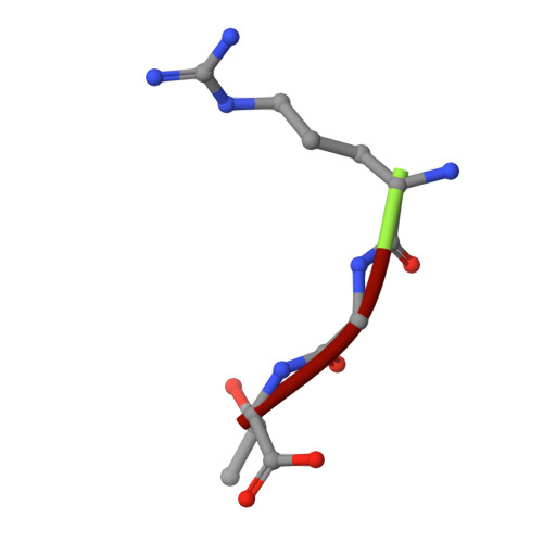
    The integrin β3-mediated c-Src priming and activation, via the SH3 domain, is consistently associated with diseases, such as the formation of thrombosis and the migration of tumor cells. Conventionally, activation of c-Src is often induced by the binding of proline-rich sequences to its SH3 domain. Instead, integrin β3 uses R(760)GT(762) for priming and activation. Because of the lack of structural information, it is not clear where RGT will bind to SH3, and under what mechanism this interaction can prime/activate c-Src. In this study, we present a 2.0-Å x-ray crystal structure in which SH3 is complexed with the RGT peptide. The binding site lies in the "N"-Src loop of the SH3 domain. Structure-based site-directed mutagenesis showed that perturbation on the "N"-Src loop disrupts the interaction between the SH3 domain and the RGT peptide. Furthermore, the simulated c-Src:β3 complex based on the crystal structure of SH3:RGT suggests that the binding of the RGT peptide might disrupt the intramolecular interaction between the SH3 and linker domains, leading to the disengagement of Trp260:"C"-helix and further activation of c-Src.


  • Organizational Affiliation

    State Key Laboratory for Medical Genomics, Shanghai Institute of Hematology, Rui Jin Hospital Affiliated to Shanghai Jiao Tong University School of Medicine, Shanghai, People's Republic of China.


Macromolecules
Find similar proteins by:  (by identity cutoff)  |  3D Structure
Entity ID: 1
MoleculeChains Sequence LengthOrganismDetailsImage
Proto-oncogene tyrosine-protein kinase Src
A, B
60Homo sapiensMutation(s): 0 
Gene Names: SRCSRC1
EC: 2.7.10.2
UniProt & NIH Common Fund Data Resources
Find proteins for P12931 (Homo sapiens)
Explore P12931 
Go to UniProtKB:  P12931
PHAROS:  P12931
GTEx:  ENSG00000197122 
Entity Groups  
Sequence Clusters30% Identity50% Identity70% Identity90% Identity95% Identity100% Identity
UniProt GroupP12931
Sequence Annotations
Expand
  • Reference Sequence

Find similar proteins by:  Sequence   |   3D Structure  

Entity ID: 2
MoleculeChains Sequence LengthOrganismDetailsImage
C-terminal 3-mer peptide from Integrin beta-33Homo sapiensMutation(s): 0 
UniProt & NIH Common Fund Data Resources
Find proteins for P05106 (Homo sapiens)
Explore P05106 
Go to UniProtKB:  P05106
PHAROS:  P05106
GTEx:  ENSG00000259207 
Entity Groups  
UniProt GroupP05106
Sequence Annotations
Expand
  • Reference Sequence
Experimental Data & Validation

Experimental Data

  • Method: X-RAY DIFFRACTION
  • Resolution: 2.00 Å
  • R-Value Free: 0.193 
  • R-Value Work: 0.139 
  • R-Value Observed: 0.144 
  • Space Group: P 1 21 1
Unit Cell:
Length ( Å )Angle ( ˚ )
a = 27.98α = 90
b = 40.83β = 105.97
c = 43.34γ = 90
Software Package:
Software NamePurpose
DNAdata collection
PHASERphasing
PHENIXrefinement
MOSFLMdata reduction
SCALAdata scaling

Structure Validation

View Full Validation Report


Entry History 

Deposition Data

Revision History  (Full details and data files)

  • Version 1.0: 2012-11-28
    Type: Initial release
  • Version 1.1: 2012-12-05
    Changes: Database references
  • Version 1.2: 2014-03-19
    Changes: Database references
  • Version 1.3: 2023-11-08
    Changes: Data collection, Database references, Refinement description