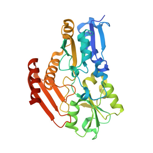
Insights into the mechanism of pyrrole polymerization catalysed by porphobilinogen deaminase: high-resolution X-ray studies of the Arabidopsis thaliana enzyme.
Roberts, A., Gill, R., Hussey, R.J., Mikolajek, H., Erskine, P.T., Cooper, J.B., Wood, S.P., Chrystal, E.J., Shoolingin-Jordan, P.M.(2013) Acta Crystallogr D Biol Crystallogr 69: 471-485
- PubMed: 23519422
- DOI: https://doi.org/10.1107/S0907444912052134
- Primary Citation of Related Structures:
4HTG - PubMed Abstract:
The enzyme porphobilinogen deaminase (PBGD; hydroxymethylbilane synthase; EC 2.5.1.61) catalyses a key early step of the haem- and chlorophyll-biosynthesis pathways in which four molecules of the monopyrrole porphobilinogen are condensed to form a linear tetrapyrrole. The active site possesses an unusual dipyrromethane cofactor which is extended during the reaction by the sequential addition of the four substrate molecules. The cofactor is linked covalently to the enzyme through a thioether bridge to the invariant Cys254. Until recently, structural data have only been available for the Escherichia coli and human forms of the enzyme. The expression of a codon-optimized gene for PBGD from Arabidopsis thaliana (thale cress) has permitted for the first time the X-ray analysis of the enzyme from a higher plant species at 1.45 Å resolution. The A. thaliana structure differs appreciably from the E. coli and human forms of the enzyme in that the active site is shielded by an extensive well defined loop region (residues 60-70) formed by highly conserved residues. This loop is completely disordered and uncharacterized in the E. coli and human PBGD structures. The new structure establishes that the dipyrromethane cofactor of the enzyme has become oxidized to the dipyrromethenone form, with both pyrrole groups approximately coplanar. Modelling of an intermediate of the elongation process into the active site suggests that the interactions observed between the two pyrrole rings of the cofactor and the active-site residues are highly specific and are most likely to represent the catalytically relevant binding mode. During the elongation cycle, it is thought that domain movements cause the bound cofactor and polypyrrole intermediates to move past the catalytic machinery in a stepwise manner, thus permitting the binding of additional substrate moieties and completion of the tetrapyrrole product. Such a model would allow the condensation reactions to be driven by the extensive interactions that are observed between the enzyme and the dipyrromethane cofactor, coupled with acid-base catalysis provided by the invariant aspartate residue Asp95.
Organizational Affiliation:
School of Biological Sciences, University of Southampton, Southampton SO16 1BJ, England.