4HSI

Glycoprotein B from Herpes simplex virus type 1, A504P/R505G/Q507G/N511G mutant, low-pH


Experimental Data Snapshot

  • Method: X-RAY DIFFRACTION
  • Resolution: 3.10 Å
  • R-Value Free: 0.255 
  • R-Value Work: 0.205 
  • R-Value Observed: 0.230 

wwPDB Validation   3D Report Full Report


Ligand Structure Quality Assessment 


This is version 2.1 of the entry. See complete history


Literature

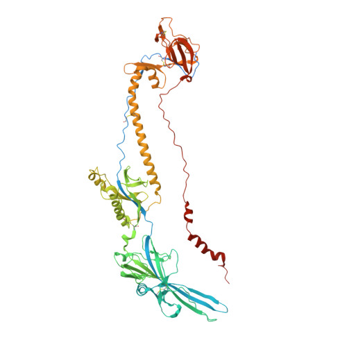
Extensive Mutagenesis of the HSV-1 gB Ectodomain Reveals Remarkable Stability of Its Postfusion Form.

Vitu, E.Sharma, S.Stampfer, S.D.Heldwein, E.E.

(2013) J Mol Biol 425: 2056-2071

  • DOI: https://doi.org/10.1016/j.jmb.2013.03.001
  • Primary Citation of Related Structures:  
    4HSI

  • PubMed Abstract: 

    Viral fusogens mediate the merger of the viral envelope and cellular membrane during viral entry. These proteins share little sequence similarity but all are thought to act by refolding through a series of conformational intermediates from the metastable prefusion form to the stable postfusion form. Crystal structures of both prefusion and postfusion forms have illuminated the conformational pathways of several viral fusogens. By contrast, only the structure of the postfusion form is available for glycoprotein B (gB), the conserved fusogen of herpesviruses. To gain insight into the nature of the fusogenic conformational changes in gB, we used several approaches aimed at engineering the prefusion form of the herpes simplex virus type 1 gB ectodomain, including modifications intended to stabilize the prefusion form and novel mutations aimed at destabilizing the postfusion form. We found that the postfusion conformation of gB is remarkably stable and resistant to perturbations. Several mutations successfully destabilized the gB trimer, identifying regions that are critical for the stability of the postfusion form. Yet, none of the constructs adopted the prefusion conformation. We propose that the soluble ectodomain of gB folds into the postfusion form without first adopting the prefusion intermediate. These results suggest that other regions of gB, including the transmembrane region and the cytoplasmic domain, may be necessary to establish and maintain the metastable prefusion conformation.


  • Organizational Affiliation

    Department of Molecular Biology and Microbiology, Tufts University School of Medicine, Boston, MA 02111, USA.


Macromolecules
Find similar proteins by:  (by identity cutoff)  |  3D Structure
Entity ID: 1
MoleculeChains Sequence LengthOrganismDetailsImage
Envelope glycoprotein B
A, B, C, D
703Human alphaherpesvirus 1 strain KOSMutation(s): 4 
Gene Names: gBUL27
UniProt
Find proteins for P06437 (Human herpesvirus 1 (strain KOS))
Explore P06437 
Go to UniProtKB:  P06437
Entity Groups  
Sequence Clusters30% Identity50% Identity70% Identity90% Identity95% Identity100% Identity
UniProt GroupP06437
Sequence Annotations
Expand
  • Reference Sequence
Oligosaccharides

Help

Entity ID: 2
MoleculeChains Length2D Diagram Glycosylation3D Interactions
2-acetamido-2-deoxy-beta-D-glucopyranose-(1-4)-2-acetamido-2-deoxy-beta-D-glucopyranose
E, F
2N-Glycosylation
Glycosylation Resources
GlyTouCan:  G42666HT
GlyCosmos:  G42666HT
GlyGen:  G42666HT
Small Molecules
Ligands 3 Unique
IDChains Name / Formula / InChI Key2D Diagram3D Interactions
NAG
Query on NAG

Download Ideal Coordinates CCD File 
G [auth A]
J [auth B]
K [auth B]
M [auth C]
P [auth D]
G [auth A],
J [auth B],
K [auth B],
M [auth C],
P [auth D],
Q [auth D]
2-acetamido-2-deoxy-beta-D-glucopyranose
C8 H15 N O6
OVRNDRQMDRJTHS-FMDGEEDCSA-N
MRY
Query on MRY

Download Ideal Coordinates CCD File 
H [auth A],
N [auth C]
MESO-ERYTHRITOL
C4 H10 O4
UNXHWFMMPAWVPI-ZXZARUISSA-N
CL
Query on CL

Download Ideal Coordinates CCD File 
I [auth A],
L [auth B],
O [auth C],
R [auth D]
CHLORIDE ION
Cl
VEXZGXHMUGYJMC-UHFFFAOYSA-M
Experimental Data & Validation

Experimental Data

  • Method: X-RAY DIFFRACTION
  • Resolution: 3.10 Å
  • R-Value Free: 0.255 
  • R-Value Work: 0.205 
  • R-Value Observed: 0.230 
  • Space Group: P 3
Unit Cell:
Length ( Å )Angle ( ˚ )
a = 117.295α = 90
b = 117.295β = 90
c = 321.872γ = 120
Software Package:
Software NamePurpose
HKL-2000data collection
PHENIXmodel building
PHENIXrefinement
HKL-2000data reduction
HKL-2000data scaling
PHENIXphasing

Structure Validation

View Full Validation Report


Ligand Structure Quality Assessment 


Entry History 

Deposition Data

Revision History  (Full details and data files)

  • Version 1.0: 2013-03-27
    Type: Initial release
  • Version 1.1: 2013-05-29
    Changes: Database references
  • Version 2.0: 2020-07-29
    Type: Remediation
    Reason: Carbohydrate remediation
    Changes: Advisory, Atomic model, Data collection, Database references, Derived calculations, Structure summary
  • Version 2.1: 2023-09-20
    Changes: Data collection, Database references, Refinement description, Structure summary