4HQX

CRYSTAL STRUCTURE OF HUMAN PDGF-BB IN COMPLEX WITH A Modified nucleotide aptamer (SOMAmer SL4)


Experimental Data Snapshot

  • Method: X-RAY DIFFRACTION
  • Resolution: 2.30 Å
  • R-Value Free: 0.279 
  • R-Value Work: 0.239 
  • R-Value Observed: 0.241 

wwPDB Validation   3D Report Full Report


This is version 1.2 of the entry. See complete history


Literature

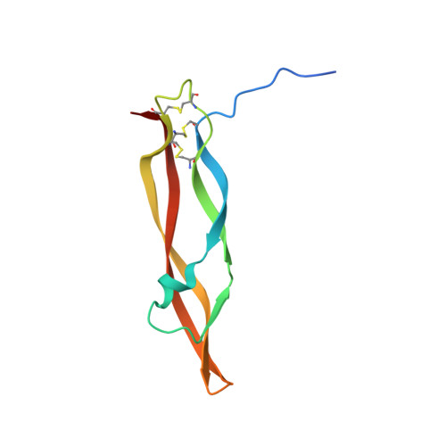
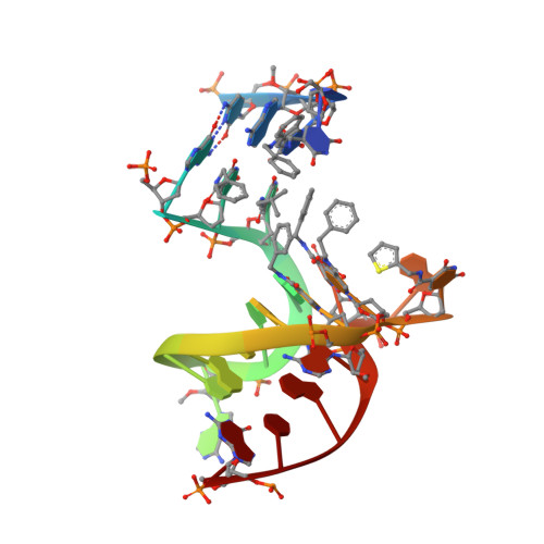
Unique motifs and hydrophobic interactions shape the binding of modified DNA ligands to protein targets.

Davies, D.R.Gelinas, A.D.Zhang, C.Rohloff, J.C.Carter, J.D.O'Connell, D.Waugh, S.M.Wolk, S.K.Mayfield, W.S.Burgin, A.B.Edwards, T.E.Stewart, L.J.Gold, L.Janjic, N.Jarvis, T.C.

(2012) Proc Natl Acad Sci U S A 109: 19971-19976

  • DOI: https://doi.org/10.1073/pnas.1213933109
  • Primary Citation of Related Structures:  
    4HQU, 4HQX

  • PubMed Abstract: 

    Selection of aptamers from nucleic acid libraries by in vitro evolution represents a powerful method of identifying high-affinity ligands for a broad range of molecular targets. Nevertheless, a sizeable fraction of proteins remain difficult targets due to inherently limited chemical diversity of nucleic acids. We have exploited synthetic nucleotide modifications that confer protein-like diversity on a nucleic acid scaffold, resulting in a new generation of binding reagents called SOMAmers (Slow Off-rate Modified Aptamers). Here we report a unique crystal structure of a SOMAmer bound to its target, platelet-derived growth factor B (PDGF-BB). The SOMAmer folds into a compact structure and exhibits a hydrophobic binding surface that mimics the interface between PDGF-BB and its receptor, contrasting sharply with mainly polar interactions seen in traditional protein-binding aptamers. The modified nucleotides circumvent the intrinsic diversity constraints of natural nucleic acids, thereby greatly expanding the structural vocabulary of nucleic acid ligands and considerably broadening the range of accessible protein targets.


  • Organizational Affiliation

    Emerald BioStructures, Inc, Bainbridge Island, WA 98110, USA.


Macromolecules

Find similar proteins by:  (by identity cutoff)  |  3D Structure
Entity ID: 1
MoleculeChains Sequence LengthOrganismDetailsImage
Platelet-derived growth factor subunit B102Homo sapiensMutation(s): 0 
UniProt & NIH Common Fund Data Resources
Find proteins for P01127 (Homo sapiens)
Explore P01127 
Go to UniProtKB:  P01127
PHAROS:  P01127
GTEx:  ENSG00000100311 
Entity Groups  
Sequence Clusters30% Identity50% Identity70% Identity90% Identity95% Identity100% Identity
UniProt GroupP01127
Sequence Annotations
Expand
  • Reference Sequence

Find similar nucleic acids by:  Sequence   |   3D Structure  

Entity ID: 2
MoleculeChains LengthOrganismImage
SOMAmer SL4B [auth C]24N/A
Sequence Annotations
Expand
  • Reference Sequence
Small Molecules
Experimental Data & Validation

Experimental Data

  • Method: X-RAY DIFFRACTION
  • Resolution: 2.30 Å
  • R-Value Free: 0.279 
  • R-Value Work: 0.239 
  • R-Value Observed: 0.241 
  • Space Group: P 41 21 2
Unit Cell:
Length ( Å )Angle ( ˚ )
a = 59.43α = 90
b = 59.43β = 90
c = 168.16γ = 90
Software Package:
Software NamePurpose
PHASERphasing
REFMACrefinement

Structure Validation

View Full Validation Report


Entry History 

Deposition Data

Revision History  (Full details and data files)

  • Version 1.0: 2012-11-21
    Type: Initial release
  • Version 1.1: 2012-12-19
    Changes: Database references
  • Version 1.2: 2023-09-20
    Changes: Advisory, Data collection, Database references, Derived calculations, Refinement description