4H6J

Identification of Cys 255 in HIF-1 as a novel site for development of covalent inhibitors of HIF-1 /ARNT PasB domain protein-protein interaction.


Experimental Data Snapshot

  • Method: X-RAY DIFFRACTION
  • Resolution: 1.52 Å
  • R-Value Free: 0.248 
  • R-Value Work: 0.215 
  • R-Value Observed: 0.216 

wwPDB Validation   3D Report Full Report


This is version 1.3 of the entry. See complete history


Literature

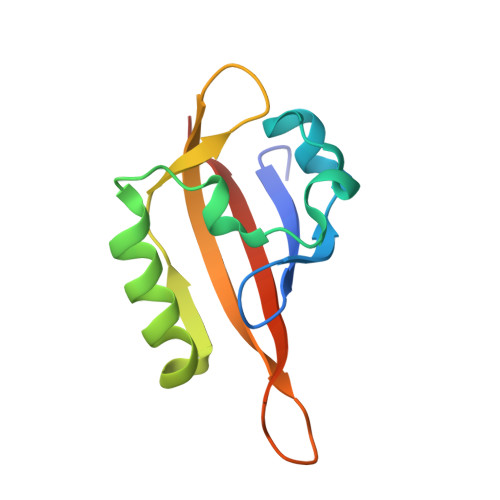
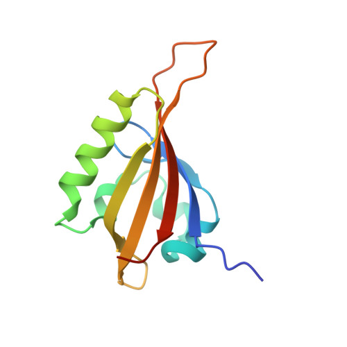
Identification of Cys255 in HIF-1 alpha as a novel site for development of covalent inhibitors of HIF-1 alpha /ARNT PasB domain protein-protein interaction.

Cardoso, R.Love, R.Nilsson, C.L.Bergqvist, S.Nowlin, D.Yan, J.Liu, K.K.Zhu, J.Chen, P.Deng, Y.L.Dyson, H.J.Greig, M.J.Brooun, A.

(2012) Protein Sci 21: 1885-1896

  • DOI: https://doi.org/10.1002/pro.2172
  • Primary Citation of Related Structures:  
    4H6J

  • PubMed Abstract: 

    The heterodimer HIF-1α (hypoxia inducible factor)/HIF-β (also known as ARNT-aryl hydrocarbon nuclear translocator) is a key mediator of cellular response to hypoxia. The interaction between these monomer units can be modified by the action of small molecules in the binding interface between their C-terminal heterodimerization (PasB) domains. Taking advantage of the presence of several cysteine residues located in the allosteric cavity of HIF-1α PasB domain, we applied a cysteine-based reactomics "hotspot identification" strategy to locate regions of HIF-1α PasB domain critical for its interaction with ARNT. COMPOUND 5 was identified using a mass spectrometry-based primary screening strategy and was shown to react specifically with Cys255 of the HIF-1α PasB domain. Biophysical characterization of the interaction between PasB domains of HIF-1α and ARNT revealed that covalent binding of COMPOUND 5 to Cys255 reduced binding affinity between HIF-1α and ARNT PasB domains approximately 10-fold. Detailed NMR structural analysis of HIF-1α-PasB-COMPOUND 5 conjugate showed significant local conformation changes in the HIF-1α associated with key residues involved in the HIF-1α/ARNT PasB domain interaction as revealed by the crystal structure of the HIF-1α/ARNT PasB heterodimer. Our screening strategy could be applied to other targets to identify pockets surrounding reactive cysteines suitable for development of small molecule modulators of protein function.


  • Organizational Affiliation

    Oncology Chemistry, Worldwide Research and Development, Pfizer Inc., San Diego, California 92121, USA.


Macromolecules
Find similar proteins by:  (by identity cutoff)  |  3D Structure
Entity ID: 1
MoleculeChains Sequence LengthOrganismDetailsImage
HYPOXIA INDUCIBLE FACTOR 1-ALPHA113Homo sapiensMutation(s): 1 
Gene Names: HIF-1 alpha
UniProt & NIH Common Fund Data Resources
Find proteins for Q16665 (Homo sapiens)
Explore Q16665 
Go to UniProtKB:  Q16665
PHAROS:  Q16665
GTEx:  ENSG00000100644 
Entity Groups  
Sequence Clusters30% Identity50% Identity70% Identity90% Identity95% Identity100% Identity
UniProt GroupQ16665
Sequence Annotations
Expand
  • Reference Sequence
Find similar proteins by:  (by identity cutoff)  |  3D Structure
Entity ID: 2
MoleculeChains Sequence LengthOrganismDetailsImage
ARYL HYDROCARBON NUCLEAR TRANSLOCATOR116Homo sapiensMutation(s): 1 
Gene Names: ARNT
UniProt & NIH Common Fund Data Resources
Find proteins for P27540 (Homo sapiens)
Explore P27540 
Go to UniProtKB:  P27540
PHAROS:  P27540
GTEx:  ENSG00000143437 
Entity Groups  
Sequence Clusters30% Identity50% Identity70% Identity90% Identity95% Identity100% Identity
UniProt GroupP27540
Sequence Annotations
Expand
  • Reference Sequence
Experimental Data & Validation

Experimental Data

  • Method: X-RAY DIFFRACTION
  • Resolution: 1.52 Å
  • R-Value Free: 0.248 
  • R-Value Work: 0.215 
  • R-Value Observed: 0.216 
  • Space Group: C 1 2 1
Unit Cell:
Length ( Å )Angle ( ˚ )
a = 68.841α = 90
b = 44.94β = 96.54
c = 78.087γ = 90
Software Package:
Software NamePurpose
CNSrefinement
CNXrefinement
PROCESSdata reduction
SCALAdata scaling
CNSphasing

Structure Validation

View Full Validation Report


Entry History 

Deposition Data

Revision History  (Full details and data files)

  • Version 1.0: 2012-12-05
    Type: Initial release
  • Version 1.1: 2013-08-28
    Changes: Database references
  • Version 1.2: 2018-01-24
    Changes: Structure summary
  • Version 1.3: 2024-02-28
    Changes: Data collection, Database references