4GUL

Study on structure and function relationships in human ferric Pirin


Experimental Data Snapshot

  • Method: X-RAY DIFFRACTION
  • Resolution: 1.80 Å
  • R-Value Free: 0.234 
  • R-Value Work: 0.187 
  • R-Value Observed: 0.190 

wwPDB Validation   3D Report Full Report


This is version 1.3 of the entry. See complete history


Literature

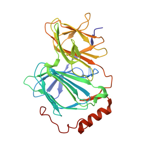
Pirin is an iron-dependent redox regulator of NF-kappa B.

Liu, F.Rehmani, I.Esaki, S.Fu, R.Chen, L.de Serrano, V.Liu, A.

(2013) Proc Natl Acad Sci U S A 110: 9722-9727

  • DOI: https://doi.org/10.1073/pnas.1221743110
  • Primary Citation of Related Structures:  
    4ERO, 4EWA, 4EWD, 4EWE, 4GUL, 4HLT

  • PubMed Abstract: 

    Pirin is a nuclear nonheme Fe protein of unknown function present in all human tissues. Here we describe that pirin may act as a redox sensor for the nuclear factor κB (NF-κB) transcription factor, a critical mediator of intracellular signaling that has been linked to cellular responses to proinflammatory signals and controls the expression of a vast array of genes involved in immune and stress responses. Pirin's regulatory effect was tested with several metals and at different oxidations states, and our spectroscopic results show that only the ferric form of pirin substantially facilitates binding of NF-κB proteins to target κB genes, a finding that suggests that pirin performs a redox-sensing role in NF-κB regulation. The molecular mechanism of such a metal identity- and redox state-dependent regulation is revealed by our structural studies of pirin. The ferrous and ferric pirin proteins differ only by one electron, yet they have distinct conformations. The Fe center is shown to play an allosteric role on an R-shaped surface area that has two distinct conformations based on the identity and the formal redox state of the metal. We show that the R-shaped area composes the interface for pirin-NF-κB binding that is responsible for modulation of NF-κB's DNA-binding properties. The nonheme Fe protein pirin is proposed to serve as a reversible functional switch that enables NF-κB to respond to changes in the redox levels of the cell nucleus.


  • Organizational Affiliation

    Department of Chemistry and Center for Diagnostics and Therapeutics, Georgia State University, Atlanta, GA 30303, USA.


Macromolecules
Find similar proteins by:  (by identity cutoff)  |  3D Structure
Entity ID: 1
MoleculeChains Sequence LengthOrganismDetailsImage
Pirin288Homo sapiensMutation(s): 0 
Gene Names: humanPIR
EC: 1.13.11.24
UniProt & NIH Common Fund Data Resources
Find proteins for O00625 (Homo sapiens)
Explore O00625 
Go to UniProtKB:  O00625
PHAROS:  O00625
GTEx:  ENSG00000087842 
Entity Groups  
Sequence Clusters30% Identity50% Identity70% Identity90% Identity95% Identity100% Identity
UniProt GroupO00625
Sequence Annotations
Expand
  • Reference Sequence
Small Molecules
Ligands 1 Unique
IDChains Name / Formula / InChI Key2D Diagram3D Interactions
FE
Query on FE

Download Ideal Coordinates CCD File 
B [auth A]FE (III) ION
Fe
VTLYFUHAOXGGBS-UHFFFAOYSA-N
Experimental Data & Validation

Experimental Data

  • Method: X-RAY DIFFRACTION
  • Resolution: 1.80 Å
  • R-Value Free: 0.234 
  • R-Value Work: 0.187 
  • R-Value Observed: 0.190 
  • Space Group: P 21 21 21
Unit Cell:
Length ( Å )Angle ( ˚ )
a = 42.305α = 90
b = 67.031β = 90
c = 107.058γ = 90
Software Package:
Software NamePurpose
SCALEPACKdata scaling
REFMACrefinement
PDB_EXTRACTdata extraction
CrystalCleardata collection
HKL-2000data reduction
HKL-2000data scaling
MOLREPphasing

Structure Validation

View Full Validation Report


Entry History 

Deposition Data

Revision History  (Full details and data files)

  • Version 1.0: 2013-05-29
    Type: Initial release
  • Version 1.1: 2013-08-28
    Changes: Database references
  • Version 1.2: 2016-12-07
    Changes: Non-polymer description
  • Version 1.3: 2023-09-13
    Changes: Data collection, Database references, Derived calculations, Refinement description