4G54

Crystal structure of a periplasmic domain of the EpsAB fusion protein of the Vibrio vulnificus type II secretion system


Experimental Data Snapshot

  • Method: X-RAY DIFFRACTION
  • Resolution: 1.55 Å
  • R-Value Free: 0.202 
  • R-Value Work: 0.175 
  • R-Value Observed: 0.176 

wwPDB Validation   3D Report Full Report


This is version 1.1 of the entry. See complete history


Literature

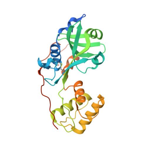
Structure of a periplasmic domain of the EpsAB fusion protein of the Vibrio vulnificus type II secretion system.

Martynowski, D.Grochulski, P.Howard, P.S.

(2013) Acta Crystallogr D Biol Crystallogr 69: 142-149

  • DOI: https://doi.org/10.1107/S0907444912042710
  • Primary Citation of Related Structures:  
    4G54

  • PubMed Abstract: 

    Vibrio vulnificus utilizes the type II secretion system (T2SS), culminating in a megadalton outer membrane complex called the secretin, to translocate extracellular proteins from the periplasmic space across the outer membrane. In Aeromonas hydrophila, the general secretion pathway proteins ExeA and ExeB form an inner membrane complex which interacts with peptidoglycan and is required for the assembly of the secretin composed of ExeD. In V. vulnificus, these two proteins are fused into one protein, EpsAB. Here, the crystal structure of a periplasmic domain of EpsAB (amino acids 333-584) solved by SAD phasing is presented. The crystals belonged to space group C2 and diffracted to 1.55 Å resolution.


  • Organizational Affiliation

    Department of Microbiology and Immunology, University of Saskatchewan, 107 Wiggins Road, Saskatoon, SK S7N 2W8, Canada.


Macromolecules
Find similar proteins by:  (by identity cutoff)  |  3D Structure
Entity ID: 1
MoleculeChains Sequence LengthOrganismDetailsImage
General Secretion Pathway Protein258Vibrio vulnificusMutation(s): 0 
Entity Groups  
Sequence Clusters30% Identity50% Identity70% Identity90% Identity95% Identity100% Identity
Sequence Annotations
Expand
  • Reference Sequence
Experimental Data & Validation

Experimental Data

  • Method: X-RAY DIFFRACTION
  • Resolution: 1.55 Å
  • R-Value Free: 0.202 
  • R-Value Work: 0.175 
  • R-Value Observed: 0.176 
  • Space Group: C 1 2 1
Unit Cell:
Length ( Å )Angle ( ˚ )
a = 128.821α = 90
b = 44.523β = 110.59
c = 51.04γ = 90
Software Package:
Software NamePurpose
MsDCdata collection
PHENIXmodel building
REFMACrefinement
HKL-2000data reduction
HKL-2000data scaling
PHENIXphasing

Structure Validation

View Full Validation Report


Entry History 

Deposition Data

Revision History  (Full details and data files)

  • Version 1.0: 2013-01-30
    Type: Initial release
  • Version 1.1: 2013-03-27
    Changes: Database references