4FMG

Merkel Cell Polyomavirus VP1 Unassembled Pentamer


Experimental Data Snapshot

  • Method: X-RAY DIFFRACTION
  • Resolution: 2.10 Å
  • R-Value Free: 0.227 
  • R-Value Work: 0.182 
  • R-Value Observed: 0.183 

wwPDB Validation   3D Report Full Report


This is version 1.1 of the entry. See complete history


Literature

Structures of Merkel Cell Polyomavirus VP1 Complexes Define a Sialic Acid Binding Site Required for Infection.

Neu, U.Hengel, H.Blaum, B.S.Schowalter, R.M.Macejak, D.Gilbert, M.Wakarchuk, W.W.Imamura, A.Ando, H.Kiso, M.Arnberg, N.Garcea, R.L.Peters, T.Buck, C.B.Stehle, T.

(2012) PLoS Pathog 8: e1002738-e1002738

  • DOI: https://doi.org/10.1371/journal.ppat.1002738
  • Primary Citation of Related Structures:  
    4FMG, 4FMH

  • PubMed Abstract: 

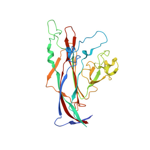
    The recently discovered human Merkel cell polyomavirus (MCPyV or MCV) causes the aggressive Merkel cell carcinoma (MCC) in the skin of immunocompromised individuals. Conflicting reports suggest that cellular glycans containing sialic acid (Neu5Ac) may play a role in MCPyV infectious entry. To address this question, we solved X-ray structures of the MCPyV major capsid protein VP1 both alone and in complex with several sialylated oligosaccharides. A shallow binding site on the apical surface of the VP1 capsomer recognizes the disaccharide Neu5Ac-α2,3-Gal through a complex network of interactions. MCPyV engages Neu5Ac in an orientation and with contacts that differ markedly from those observed in other polyomavirus complexes with sialylated receptors. Mutations in the Neu5Ac binding site abolish MCPyV infection, highlighting the relevance of the Neu5Ac interaction for MCPyV entry. Our study thus provides a powerful platform for the development of MCPyV-specific vaccines and antivirals. Interestingly, engagement of sialic acid does not interfere with initial attachment of MCPyV to cells, consistent with a previous proposal that attachment is mediated by a class of non-sialylated carbohydrates called glycosaminoglycans. Our results therefore suggest a model in which sialylated glycans serve as secondary, post-attachment co-receptors during MCPyV infectious entry. Since cell-surface glycans typically serve as primary attachment receptors for many viruses, we identify here a new role for glycans in mediating, and perhaps even modulating, post-attachment entry processes.


  • Organizational Affiliation

    Interfaculty Institute of Biochemistry, University of Tuebingen, Tuebingen, Germany.


Macromolecules
Find similar proteins by:  (by identity cutoff)  |  3D Structure
Entity ID: 1
MoleculeChains Sequence LengthOrganismDetailsImage
VP1
A, B, C, D, E
A, B, C, D, E, F, G, H, I, J, K, L, M, N, O, P, Q, R, S, T
289Merkel cell polyomavirusMutation(s): 0 
Gene Names: VP1
UniProt
Find proteins for C0JPK1 (Merkel cell polyomavirus)
Explore C0JPK1 
Go to UniProtKB:  C0JPK1
Entity Groups  
Sequence Clusters30% Identity50% Identity70% Identity90% Identity95% Identity100% Identity
UniProt GroupC0JPK1
Sequence Annotations
Expand
  • Reference Sequence
Small Molecules
Ligands 2 Unique
IDChains Name / Formula / InChI Key2D Diagram3D Interactions
GOL
Query on GOL

Download Ideal Coordinates CCD File 
AB [auth L]
BA [auth C]
BB [auth L]
CA [auth C]
DB [auth M]
AB [auth L],
BA [auth C],
BB [auth L],
CA [auth C],
DB [auth M],
EA [auth D],
FA [auth D],
FB [auth N],
HA [auth E],
HB [auth O],
JA [auth F],
JB [auth P],
KA [auth F],
LB [auth Q],
MA [auth G],
NB [auth R],
OA [auth H],
PA [auth H],
PB [auth S],
QB [auth S],
RA [auth I],
SA [auth I],
SB [auth T],
U [auth A],
UA [auth J],
V [auth A],
VA [auth J],
W [auth A],
XA [auth K],
Y [auth B],
Z [auth B],
ZA [auth L]
GLYCEROL
C3 H8 O3
PEDCQBHIVMGVHV-UHFFFAOYSA-N
CL
Query on CL

Download Ideal Coordinates CCD File 
AA [auth B]
CB [auth L]
DA [auth C]
EB [auth M]
GA [auth D]
AA [auth B],
CB [auth L],
DA [auth C],
EB [auth M],
GA [auth D],
GB [auth N],
IA [auth E],
IB [auth O],
KB [auth P],
LA [auth F],
MB [auth Q],
NA [auth G],
OB [auth R],
QA [auth H],
RB [auth S],
TA [auth I],
TB [auth T],
WA [auth J],
X [auth A],
YA [auth K]
CHLORIDE ION
Cl
VEXZGXHMUGYJMC-UHFFFAOYSA-M
Experimental Data & Validation

Experimental Data

  • Method: X-RAY DIFFRACTION
  • Resolution: 2.10 Å
  • R-Value Free: 0.227 
  • R-Value Work: 0.182 
  • R-Value Observed: 0.183 
  • Space Group: P 1
Unit Cell:
Length ( Å )Angle ( ˚ )
a = 85.27α = 92.98
b = 85.62β = 100.5
c = 248.17γ = 108.05
Software Package:
Software NamePurpose
MxCuBEdata collection
PHASERphasing
REFMACrefinement
XDSdata reduction
XSCALEdata scaling

Structure Validation

View Full Validation Report


Entry History 

Deposition Data

Revision History  (Full details and data files)

  • Version 1.0: 2012-09-05
    Type: Initial release
  • Version 1.1: 2024-02-28
    Changes: Data collection, Database references, Derived calculations