4EYR

Crystal structure of multidrug-resistant clinical isolate 769 HIV-1 protease in complex with ritonavir


Experimental Data Snapshot

  • Method: X-RAY DIFFRACTION
  • Resolution: 1.80 Å
  • R-Value Free: 0.226 
  • R-Value Work: 0.191 
  • R-Value Observed: 0.193 

wwPDB Validation   3D Report Full Report


Ligand Structure Quality Assessment 


This is version 1.3 of the entry. See complete history


Literature

Insights into the mechanism of drug resistance: X-ray structure analysis of multi-drug resistant HIV-1 protease ritonavir complex.

Liu, Z.Yedidi, R.S.Wang, Y.Dewdney, T.G.Reiter, S.J.Brunzelle, J.S.Kovari, I.A.Kovari, L.C.

(2013) Biochem Biophys Res Commun 431: 232-238

  • DOI: https://doi.org/10.1016/j.bbrc.2012.12.127
  • Primary Citation of Related Structures:  
    4EYR

  • PubMed Abstract: 

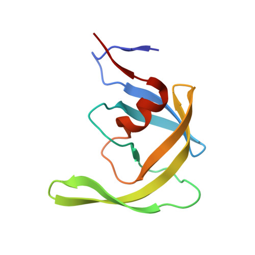
    Ritonavir (RTV) is a first generation HIV-1 protease inhibitor with rapidly emerging drug resistance. Mutations at residues 46, 54, 82 and 84 render the HIV-1 protease drug resistant against RTV. We report the crystal structure of multi-drug resistant (MDR) 769 HIV-1 protease (carrying resistant mutations at residues 10, 36, 46, 54, 62, 63, 71, 82, 84 and 90) complexed with RTV and the in vitro enzymatic IC(50) of RTV against MDR HIV-1 protease. The structural and functional studies demonstrate significant drug resistance of MDR HIV-1 protease against RTV, arising from reduced hydrogen bonds and Van der Waals interactions between RTV and MDR HIV-1 protease.


  • Organizational Affiliation

    Department of Biochemistry and Molecular Biology, School of Medicine, Wayne State University, Detroit, MI 48201, USA.


Macromolecules
Find similar proteins by:  (by identity cutoff)  |  3D Structure
Entity ID: 1
MoleculeChains Sequence LengthOrganismDetailsImage
HIV-1 PROTEASE
A, B
99Human immunodeficiency virus 1Mutation(s): 4 
Gene Names: pol
UniProt
Find proteins for Q000H7 (Human immunodeficiency virus 1)
Explore Q000H7 
Go to UniProtKB:  Q000H7
Entity Groups  
Sequence Clusters30% Identity50% Identity70% Identity90% Identity95% Identity100% Identity
UniProt GroupQ000H7
Sequence Annotations
Expand
  • Reference Sequence
Small Molecules
Ligands 1 Unique
IDChains Name / Formula / InChI Key2D Diagram3D Interactions
RIT
Query on RIT

Download Ideal Coordinates CCD File 
C [auth B]RITONAVIR
C37 H48 N6 O5 S2
NCDNCNXCDXHOMX-XGKFQTDJSA-N
Binding Affinity Annotations 
IDSourceBinding Affinity
RIT BindingDB:  4EYR Ki: min: 0.01, max: 109 (nM) from 29 assay(s)
Kd: min: 0.6, max: 0.61 (nM) from 2 assay(s)
IC50: min: 67, max: 9850 (nM) from 2 assay(s)
EC50: 150 (nM) from 1 assay(s)
-TΔS: min: -1.02e+2, max: -3.01e+1 (kJ/mol) from 26 assay(s)
ΔH: min: -2.76e+1, max: 73.15 (kJ/mol) from 29 assay(s)
ΔG: min: -6.02e+1, max: -2.88e+1 (kJ/mol) from 28 assay(s)
PDBBind:  4EYR IC50: 237 (nM) from 1 assay(s)
Binding MOAD:  4EYR IC50: 237 (nM) from 1 assay(s)
Biologically Interesting Molecules (External Reference) 1 Unique
Experimental Data & Validation

Experimental Data

  • Method: X-RAY DIFFRACTION
  • Resolution: 1.80 Å
  • R-Value Free: 0.226 
  • R-Value Work: 0.191 
  • R-Value Observed: 0.193 
  • Space Group: P 41
Unit Cell:
Length ( Å )Angle ( ˚ )
a = 45.398α = 90
b = 45.398β = 90
c = 104.071γ = 90
Software Package:
Software NamePurpose
MOLREPphasing
REFMACrefinement
SCALAdata scaling

Structure Validation

View Full Validation Report


Ligand Structure Quality Assessment 


Entry History 

Deposition Data

Revision History  (Full details and data files)

  • Version 1.0: 2013-01-30
    Type: Initial release
  • Version 1.1: 2013-02-27
    Changes: Database references
  • Version 1.2: 2018-06-27
    Changes: Data collection, Database references
  • Version 1.3: 2024-02-28
    Changes: Data collection, Database references, Derived calculations