


Structure based functional analysis of Staphylococcal Phosphoglycerate kinase
Roychowdhury, A., Mukherjee, S., Dutta, D., Das, A.K.To be published.
Experimental Data Snapshot
wwPDB Validation 3D Report Full Report

Entity ID: 1 | |||||
|---|---|---|---|---|---|
| Molecule | Chains | Sequence Length | Organism | Details | Image |
| Phosphoglycerate kinase | 403 | Staphylococcus aureus subsp. aureus MRSA252 | Mutation(s): 0 Gene Names: pgk, SAR0829 EC: 2.7.2.3 | 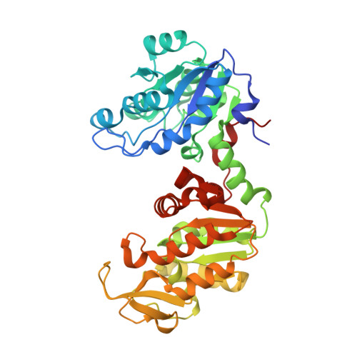 | |
UniProt | |||||
Find proteins for Q6GIL7 (Staphylococcus aureus (strain MRSA252)) Explore Q6GIL7 Go to UniProtKB: Q6GIL7 | |||||
Entity Groups | |||||
| Sequence Clusters | 30% Identity50% Identity70% Identity90% Identity95% Identity100% Identity | ||||
| UniProt Group | Q6GIL7 | ||||
Sequence AnnotationsExpand | |||||
| |||||
| Length ( Å ) | Angle ( ˚ ) |
|---|---|
| a = 45.137 | α = 90 |
| b = 74.748 | β = 95.72 |
| c = 58.665 | γ = 90 |
| Software Name | Purpose |
|---|---|
| SCALA | data scaling |
| MOLREP | phasing |
| REFMAC | refinement |
| PDB_EXTRACT | data extraction |
| StructureStudio | data collection |
| XDS | data reduction |
RCSB PDB (citation) is hosted by
RCSB PDB is a member of the

